ENZO热销产品——ER-ID® Green assay kit内质网检测试剂盒(绿色)

ENZO热销产品——ER-ID® Green assay kit内质网检测试剂盒(绿色)

ENZO热销产品——ER-ID® Green assay kit内质网检测试剂盒(绿色)


Enzo Life Sciences公司的ER-ID® Green assay kit适用于活细胞或破膜醛固定细胞的染色。微摩尔级浓度的ER-ID®绿色染料足以对哺乳动物细胞进行染色,在人宫颈癌细胞系、T淋巴细胞系、Jurkat 细胞系、HeLa 细胞系以及骨肉瘤上皮细胞U2-OS中已经得到了验证。ER-ID® Green assay kit专门用于表达RFP或OFP的细胞系,以及表达蓝色或青色荧光蛋白(BFP、CFP)的细胞。该试剂盒适用于活细胞或固定后细胞与探针(如标记抗体)或与Texas Red或香豆素类似光谱性质的其他荧光缀合物结合使用。


产品特点

● 新型内质网染料,可用于活细胞、破膜细胞或固定细胞的染色

● 与常见荧光染料和荧光蛋白兼容

● 高耐光漂白性、抗浓度淬灭和光转换

● 生产严格,以控制和消除非特异性结果


实例分析

● ER-ID®绿色染色与含有KDEL序列的钙网蛋白共定位,与橙色荧光蛋白(OFP)融合。
ENZO热销产品——ER-ID® Green assay kit内质网检测试剂盒(绿色)


● 用ER-ID®绿色染料(A)、Hoechst 33342染料(B)对HeLa活细胞进行染色,得到的合成图像(C)。

ENZO热销产品——ER-ID® Green assay kit内质网检测试剂盒(绿色)


● 用4%多聚甲醛固定HepG2细胞20分钟,并用ER-ID®标记30分钟。下图为标记3天后拍摄的照片。
ENZO热销产品——ER-ID® Green assay kit内质网检测试剂盒(绿色)


● 将HepG2细胞与ER-ID®孵育30分钟,并用倒置显微镜(Zeiss)进行分析。
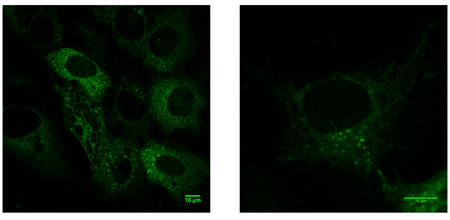
ENZO热销产品——ER-ID® Green assay kit内质网检测试剂盒(绿色)


产品详情:

产品名称

ER-ID® Green assay kit

产品货号

ENZ-51025-K500

产品规格

500 assays

质量控制

A sample from each lot of ER-ID® Green assay kit is used to stain HeLa cells, using the procedures described in the user manual. The selectivity of the ER-ID® Green dye is evident.

应用

Fluorescence microscopy

Enzo Life Sciences’ ER-ID® Green assay kit contains a novel endoplasmic reticulum-selective dye suitable for live cell, or detergent-permeabilized aldehyde-fixed cell staining.

长期储存

-20°C

组分

ER-ID® Green detection reagent: 50μl

Hoechst 33342 nuclear stain: 50μl

10X assay buffer: 15ml

产品说明

The ER-ID® Green assay kit is a member of the CELLESTIAL® product line, reagents and assay kits comprising fluorescent molecular probes that have been extensively benchmarked for live cell analysis applications. CELLESTIAL® reagents


部分产品文献引用

1. Molecular interplay between NOX1 and autophagy in cadmium-induced prostate carcinogenesis: A. Tyagi, et al.; Free Radic. Biol. Med. 199, 44 (2023) 

2. Ammonia induces amyloidogenesis in astrocytes by promoting amyloid precursor protein translocation into the endoplasmic reticulum: A. Komatsu, et al.; J. Biol. Chem. 298, 101933 (2022) 

3. PLP1 mutations in patients with multiple sclerosis: identification of a new mutation and potential pathogenicity of the mutations: N.C. Cloake, et al.; J. Clin. Med. 7, 342 (2018), Application(s): Fluorescence microscopy 

4. Englerin A induces an acute inflammatory response and reveals lipid metabolism and ER stress as targetable vulnerabilities in renal cell carcinoma: A. Batova, et al.; PLoS One 12, e0172632 (2017), Application(s): Fluorescence microscopy

5. Cell growth on (“Janus”) density gradients of bifunctional zeolite L crystals: N.S. Kehr, et al.; ACS Appl. Mater. Interfaces 8, 35081 (2016)

6. The parathyroid hormone second receptor PTH2R and its ligand tuberoinfundibular peptide of 39 residues TIP39 regulate intracellular calcium and influence keratinocyte differentiation: E. Sato, et al.; J. Invest. Dermatol. 136, 1449 (2016)

7. Viral genome imaging of hepatitis C virus to probe heterogeneous viral infection and responses to antiviral therapies: V. Ramanan, et al.; Virology 494, 236 (2016), Application(s): Endoplasmic reticulum staining

8. Localization-Dependent Cell-Killing Effects of Protoporphyrin (PPIX)-Lipid Micelles and Liposomes in Photodynamic Therapy: S. Tachikawa, et al.; Bioorg. Med. Chem. 23, 7578 (2015), Application(s): Cell staining


详情请咨询 ENZO 代理商-上海金畔生物科技

ENZO热销产品——TOTAL-NUCLEAR-ID® green/red nucleolar/nuclear detection kit核仁/细胞核检测试剂盒(绿色/红色)

ENZO热销产品——TOTAL-NUCLEAR-ID® green/red nucleolar/nuclear detection kit核仁/细胞核检测试剂盒(绿色/红色)

ENZO热销产品——TOTAL-NUCLEAR-ID® green/red nucleolar/nuclear detection kit核仁/细胞核检测试剂盒(绿色/红色)


ENZO的TOTAL-NUCLEAR-ID® green/red nucleolar/nuclear detection kit包含两种专有染料,适用于同时对细胞核和核仁进行活细胞染色。这些染料可以检验由细胞周期和核糖体发生等生物过程引起的细胞内分布、运输和定位的核仁动态变化。该试剂盒与大多数荧光检测系统兼容,包括常规和共聚焦荧光显微镜,以及高内涵筛选(HCS)平台。TOTAL-NUCLEAR-ID® green/red nucleolar/nuclear detection kit专门设计用于观察活细胞中的核仁和细胞核,潜在应用包括监测受损核糖体的生物生成、抑制转录、细胞周期动力学和细胞应激,以及核仁蛋白的分布、运输和动力学,病毒蛋白的分布,并可能有助于识别癌症细胞。


产品特点

● 同时对活细胞中的核仁和细胞核染色

● 高耐光漂白性和抗淬灭,即使在长时间观察后也能确保强烈一致的荧光信号

● 已验证在活细胞成像应用中的实用性,对核仁扰动剂的治疗有适当的反应性

● 生产严格,以控制和消除非特异性结果


实例分析

● NUCLEOLAR-ID®绿色检测试剂(A)和NUCLEOR-ID®红色检测试剂(B)的吸收和发射光谱

ENZO热销产品——TOTAL-NUCLEAR-ID® green/red nucleolar/nuclear detection kit核仁/细胞核检测试剂盒(绿色/红色)

● 用NUCLEOLAR-ID®绿色检测试剂染色的细胞在核仁内显示出最大的荧光信号,在整个细胞核和细胞质中显示出微弱的荧光(A)。当用低剂量放线菌素D处理细胞时,观察到核仁染色丢失(B)。与较弱的细胞质染色相比,使用NUCLEOLAR-ID®红色检测试剂进行反染色有助于突出核仁。

ENZO热销产品——TOTAL-NUCLEAR-ID® green/red nucleolar/nuclear detection kit核仁/细胞核检测试剂盒(绿色/红色)

● 用TOTAL-NUCLEAR-ID® Green/Red Nucleolar/ Nuclear Detection Kit对U2OS活细胞进行染色,通过荧光显微镜观察细胞核(红色)和核仁(黄绿色)的染色结果。作为一种RNA染料,NUCLEOLAR-ID®绿色染料也显示出一些细胞质染色,但NUCLEOR-ID®红色染料有助于在高亮显示的红色细胞核荧光信号范围内,将核仁明确识别为绿色荧光信号。

ENZO热销产品——TOTAL-NUCLEAR-ID® green/red nucleolar/nuclear detection kit核仁/细胞核检测试剂盒(绿色/红色) 

● U2OS细胞中染色为红色的细胞核和染色为绿色的核仁的3D重建

ENZO热销产品——TOTAL-NUCLEAR-ID® green/red nucleolar/nuclear detection kit核仁/细胞核检测试剂盒(绿色/红色)


产品详情:

产品名称 TOTAL-NUCLEAR-ID® green/red nucleolar/nuclear detection kit
产品货号 ENZ-51006-500
产品规格 500 assays
质量控制 A sample from each lot of TOTAL-NUCLEAR-ID® Green/ Red Nucleolus/Nucleus Detection Kit is used to stain HeLa cells using the procedures described in the user manual. Cells stained with NUCLEOLAR-ID® Green Detection Reagent show maximal fluorescence signal within the nucleoli, with only faint fluorescence throughout the nucleus and cytoplasm. NUCLEAR-ID® Red Detection Reagent maximally stains the DNA in the cell nucleus. Cells induced by actinomycin D show reduction of nucleolar signal.
应用 Fluorescence microscopy, Fluorescent detection
For visualizing nucleoli and nuclei simultaneously in living cells.
短期储存 -20°C
长期储存 -80°C
组分 NUCLEOLAR-ID® Green Detection Reagent, 50 µL
NUCLEAR-ID® Red Detection Reagent, 50 µL
Actinomycin D Control, 125 µg
10X Assay Buffer, 15 mL
产品说明 The TOTAL-NUCLEAR-ID® green/red nucleolar/nuclear detection kit is a member of the CELLESTIAL® product line, reagents and assay kits comprising fluorescent molecular probes that have been extensively benchmarked for live cell analysis applications. CELLESTIAL® reagents and kits are optimal for use in demanding imaging applications, such as confocal microscopy, flow cytometry and HCS, where consistency and reproducibility are required.


部分产品文献引用

1. Guanidinylated bioresponsive poly(amido amine)s designed for intranuclear gene delivery: J. Yu, et al.; Int. J. Nanomedicine 11, 4011 (2016), Application(s): Confocal microscopy using MCF7 cells

2. Dose-dependent role of the cohesin complex in normal and malignant hematopoiesis: A.D. Viny, et al.; J. Exp. Med. 212, 1819 (2015), Application(s): Confocal microscopy

3. Small-molecule multi-targeted kinase inhibitor RGB-286638 triggers P53-dependent and -independent anti-multiple myeloma activity through inhibition of transcriptional CDKs: D. Cirstea, et al.; Leukemia 27, 2366 (2013), Application(s): Nucleolar segregation analysis by immunofluorescence

4. Mismatch in mechanical and adhesive properties induces pulsating cancer cell migration in epithelial monolayer: M.H. Lee, et al.; Biophys. J. 102, 2731 (2012), Application(s): Detection of nuclei and nucleoli in breast cell lines using fluorescence microscopy

5. AFM stiffness nanotomography of normal, metaplastic and dysplastic human esophageal cells: A. Fuhrmann, et al.; Phys. Biol. 8, 015007 (2011), Application(s): Fluorescence lifetime imaging of normal and dysplasic esophageal cells

6. Strategies of actin reorganisation in plant cells: A.P. Smertenko, et al.; J. Cell Sci. 123, 3019 (2010), Application(s): Staining of plant cells


详情请咨询 ENZO 代理商-上海金畔生物科技

ENZO热销产品——EFLUXX-ID®多药耐药性分析试剂盒

ENZO热销产品——EFLUXX-ID®多药耐药性分析试剂盒

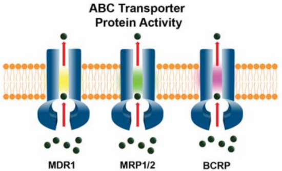
Enzo Life Sciences公司的EFLUXX-ID®多药耐药性分析试剂盒可实现对三个临床相关的ABC转运蛋白MDR1 (p-glycoprotein), MRP1/2BCRP的功能检测。试剂盒采用疏水性的非荧光化合物,很容易穿透细胞膜,细胞内脂酶将其水解为亲水性的荧光染料。除非ABC转运蛋白通过主动运输将其泵出胞外,否则产生的荧光染料将包裹在细胞内。因此如果细胞对药物有抗性作用,荧光强度就会减弱。EFLUXX-ID®多药耐药性分析试剂盒是目前少有可同时监测三种主要的ABC转运蛋白并能够分析单泵活性的试剂盒。

ENZO热销产品——EFLUXX-ID®多药耐药性分析试剂盒 


EFLUXX-ID®多药耐药性染料与其他荧光染料的兼容性

ENZO热销产品——EFLUXX-ID®多药耐药性分析试剂盒 


产品信息

货号

产品名称

规格

ENZ-51029-K100

EFLUXX-ID® Green multidrug resistance assay kit

1*1Kit(100 assays For flow cytometry)

ENZ-51030-K100

EFLUXX-ID® Gold multidrug resistance assay kit

1*1Kit(100 assays For flow cytometry)


产品特点

● 可检测与3ABC转运蛋白活性相关的多药耐药性

● 通过单一专有染料定量检测活细胞中MDR活性,并以MDR活性因子(MAF)表征

● 试剂盒内含有已知的MDR1MRP1/2BCRP特异性抑制剂

● 操作简单,无需洗涤,1h 内可获得结果

● 可检测Calcein AM无法检测到的BCRP蛋白活性

● 有绿色和金色两种荧光染料可供选择

● 两种染料均可与表达GFP的细胞系或其他CELLESTIAL®染料同时使用


实例分析

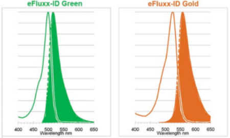
● EFLUXX-ID® Green 490/514nm ex/emEFLUXX-ID® Gold 530/570nm ex/em试剂的光谱特性可与其他常见荧光染料进行多重检测。

ENZO热销产品——EFLUXX-ID®多药耐药性分析试剂盒 


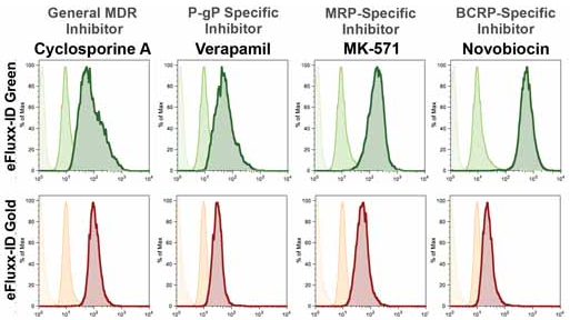
● 使用EFLUXX-ID®GreenEFLUXX-ID®Gold染料在CHO K1细胞中评估已知抑制剂对ABC转运蛋白活性的分析

ENZO热销产品——EFLUXX-ID®多药耐药性分析试剂盒 

将细胞与试剂盒中含有的MDR通用抑制剂(最左列)或转运蛋白特异性抑制剂在37°C下孵育5 min。随后在37°C下用指定的染料染色细胞30 min,并立即通过流式细胞术进行分析。所用抑制剂:5 µM Cyclosporin AMDR通用抑制剂)、20 µM VerapamilP-gp特异性抑制剂)、0.05 mM MK-571MRP特异性抑制剂)、0.05 mM NovobiocinBCRP特异性抑制剂)。


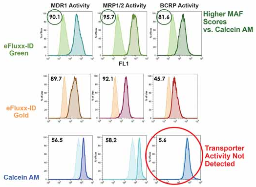
● 三种主要ABC转运蛋白的活性分析

ENZO热销产品——EFLUXX-ID®多药耐药性分析试剂盒 

 使用EFLUXX-ID®Green(上)、Gold(中)或Calcein AM(下)染料,通过流式细胞术评估CHO K1细胞中ABC转运蛋白的活性。与未处理的细胞相比,用ABC转运蛋白特异性抑制剂(图中阴影部分)处理可诱导染料在细胞内的滞留(图中实线部分)。平均荧光强度(MFI)的差异表明了对应蛋白的活性,以多药耐药性活性因子值(MAF)表征多药耐药性。结果表明EFLUXX-ID®染料对抑制剂具有良好的特异性,而Calcein AM染料(常用的MDR检测探针)无法检测BCRP活性。


部分产品文献引用

1. Overexpression of P-glycoprotein and MRP-1 are pharmacogenomic biomarkers to determine steroid resistant phenotype in childhood idiopathic nephrotic syndrome: P. Narayan, et al.; Pharmacogenomics J. 21, 566 (2021)

2. Targeting poor proteasomal function with radioiodine eliminates CT26 colon cancer stem cells resistant to bortezomib therapy: J. H. Lee, et al.; Sci. Rep. 10, 14308 (2020)

3. Class III β-Tubulin Overexpression Induces Chemoresistance to Eribulin in a Leiomyosarcoma Cell Line: K. Yahiro, et al.; Anal. Cell. Pathol. (Amst.) 2018, 8987568 (2018), Application(s): Flow cytometry. MDR1 activity in human leiomysarcoma cell line SK-LMS-1

4. Soluble uric acid increases PDZK1 and ABCG2 expression in human intestinal cell lines via the TLR4-NLRP3 inflammasome and PI3K/Akt signaling pathway: M. Chen, et al.; Arthritis Res. Ther. 20, 20 (2018)

5. ABCB1 and ABCG2 drug transporters are differentially expressed in non-small cell lung cancers (NSCLC) and expression is modified by cisplatin treatment via altered Wnt signaling: M. Vesel, et al.; Respir. Res. 18, 52 (2017)

6. Expression and activity of multidrug resistance proteins in mature endothelial cells and their precursors: A challenging correlation: A. Krawczenko, et al.; PLoS One 12, e0172371 (2017), Application(s): Comparison of different MDR detection methods in endothelial non-cancerous cells

7. Identification of volasertib-resistant mechanism and evaluation of combination effects with volasertib and other agents on acute myeloid leukemia: Y. Adachi, et al.; Oncotarget 8, 78452 (2017), Application(s): Flow Cytometry; acute myeloid leukemia

8. Isomahanine induces endoplasmic reticulum stress and simultaneously triggers p38 MAPK-mediated apoptosis and autophagy in multidrug-resistant human oral squamous cell carcinoma cells: T. Utaipan, et al.; Oncol. Rep. 37, 1243 (2017), Application(s): Flow Cytometry; oral squamous cell carcinoma (OSCC)


详情请咨询 ENZO 代理商上海金畔生物科技

全国服务热线: 15221999938            邮箱: sales@jinpanbio.com 

深圳: 021-50837765      北京: 021-50837765       上海: 021-50837765      

广州: 021-50837765        香港: 021-50837765 

代理品牌网站: www.nbs-bio.com 
自主品牌网站: www.jinpanbio.cnet

ENZO热销产品——NUCLEAR-ID®系列染料

ENZO热销产品——NUCLEAR-ID®系列染料

Enzo Life Sciences公司的NUCLEAR-ID® Blue DNA stain (GFP-CERTIFIED®)NUCLEAR-ID ® Red DNA Stain是细胞渗透性染料,适用于活细胞的细胞核染色。这系列染料为通过流式细胞术研究细胞周期进程的诱导和抑制提供了一种简便方法。该试剂在活细胞研究中的潜在应用包括确定细胞DNA含量和细胞周期分布、检测生长模式的变化、监测细胞凋亡以及评估肿瘤细胞行为和抑制基因机制。


NUCLEAR-ID® Red DNA stain


产品特点

● 远红色荧光特异性DNA染料

● 稳定且纯度高

● 可分析活细胞、可渗透细胞和固定细胞

● 无光漂白作用

● 无需RNase处理

● GFPFITC兼容

● 不需要紫外线激光源激发

● 经验证适用于各种细胞密度

● 快速且易于使


实例分析

37°C下用终浓度为4000x2000xNUCLEAR-ID ® Red DNA Stain同等µM浓度的竞争品牌染料对细胞进行染色,并在染色后轻轻洗涤。15分钟24小时细胞成像,结果显示NUCLEAR-ID ® Red DNA Stain在更低的浓度就能显现dsDNA


ENZO热销产品——NUCLEAR-ID®系列染料


产品详情:

产品名称

NUCLEAR-ID® Red DNA stain

产品货号

ENZ-52406

产品规格

200µl

最大激发波长

566nm

最大发射波长

650nm

纯度

≥93%HPLC

质量控制

1.Absorption peak of NUCLEAR-ID® Red dye: λmax = 566 ± 4 nm

2.% purity of NUCLEAR-ID® Red dye by HPLC: ≥93%

应用

Flow Cytometry, Fluorescent detection

短期储存

-20°C

长期储存

-80°C

产品说明

The NUCLEAR-ID® Red DNA Stain is a member of the CELLESTIAL® product line, reagents and assay 

kits comprising fluorescent molecular probes that have been extensively benchmarked for live cell analysis 

applications. CELLESTIAL® reagents and kits are optimal for use in demanding imaging applications, such 

as confocal microscopy, flow cytometry and HCS, where consistency and reproducibility are required.


部分产品文献引用

1. CD200R1L is a functional evolutionary conserved activating receptor in human neutrophils: M.I.P. Ramos, et al.; J. Leukoc. Biol. 111, 367 (2022)

2. FOXM1 network in association with TREM1 suppression regulates NET formation in diabetic foot ulcers: A.P. Sawaya, et al.; EMBO Rep. 23, e54558 (2022)

3. Multi-dimensional-double-spiral (MDDS) inertial microfluidic platform for sperm isolation directly from the raw semen sample: H. Jeon, et al.; Sci. Rep. 12, 4212 (2022)

4. DYRK1A negatively regulates CDK5-SOX2 pathway and self-renewal of glioblastoma stem cells: B. Chen, et al.; Int. J. Mol. Sci. 22, 4011 (2021)

5. Global phosphoproteomics reveals DYRK1A regulates CDK1 activity in glioblastoma cells: A. Recasens, et al.; Cell Death Discov. 7, 81 (2021)

6. Hydrogen Peroxide Is Crucial for NLRP3 Inflammasome-Mediated IL-1β Production and Cell Death in Pneumococcal Infections of Bronchial Epithelial Cells; Journal of Innate Immunity: S. Surabhi, et al.; J. Innate Immun. 10, 1159 (2021)

7. Licochalcone A inhibits hypoxia-inducible factor-1α accumulation by suppressing mitochondrial respiration in hypoxic cancer cells: M.K. Park, et al.; Biomed. Pharmacother. 133, 111082 (2021)


NUCLEAR-ID® Blue DNA stain (GFP-CERTIFIED®)


产品特点

● 蓝色荧光特异性DNA染料

● 稳定且纯度高

● 可分析活细胞、可渗透细胞和固定细胞

● 无光漂白作用

● 无需RNase处理

● GFP和红色染料兼容

● 经验证适用于各种细胞密度

● 快速易用


产品详情:

产品名称

NUCLEAR-ID® Blue DNA stain (GFP-CERTIFIED®)

产品货号

ENZ-CHM103-0200

产品规格

200µl

最大激发波长

350nm

最大发射波长

461nm

纯度

≥95% (HPLC)

外观

Frozen liquid

应用

Flow Cytometry, Fluorescent detection

短期储存

-20°C, Protect from light. Avoid freeze/thaw cycles

长期储存

-80°C, Protect from light. Avoid freeze/thaw cycles


详情请咨询 ENZO 代理商上海金畔生物科技

全国服务热线: 15221999938            邮箱: sales@jinpanbio.com 

深圳: 021-50837765      北京: 021-50837765       上海: 021-50837765      

广州: 021-50837765       香港: 021-50837765 

代理品牌网站: www.nbs-bio.com 
自主品牌网站: www.jinpanbio.cnet

CYTO-ID Autophagy detection kit⾃噬分析试剂盒

CYTO-ID Autophagy detection kit⾃噬分析试剂盒

CYTO-ID Autophagy detection kit⾃噬分析试剂盒–⽆需转染,流式或显微镜⽅法检测

常⽤监测细胞⾃噬的⽅式是构建LC3-GFP(绿⾊荧光蛋⽩)融合蛋⽩,LC3蛋⽩可以和⾃噬体膜发⽣特异性结合,GFP作为⽰踪剂。但是构建LC3-GFP融合基因,转染,表达需要⼀定时间,⽽且LC3-GFP表达不理想也会影响细胞⾃噬的检测。该试剂盒采⽤申请专利的⾃噬荧光检测试剂,免去转染表达的时间,特异性⾼于LC3。

CYTO-ID Autophagy detection kit⾃噬分析试剂盒

特点:

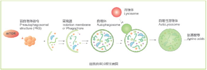
CYTO-ID Autophagy detection kit检测过程中⽆需转染,适合流式和显微镜⾼通量分析,CYTO-ID Autophagy detection kit组分包括,灵敏的荧光探针,可以染前⾃噬体,⾃噬⼩体,很少染上溶酶体。

专利的染⾃噬囊泡,⽆需耗时融合转染LC3

在多种细胞中快速定量⾃噬

能够忽略溶酶体的染⾊,降低了背景⼲扰

试剂盒中提供的成分:

Cyto-ID® 绿⾊检测试剂⾃噬检测试剂

Hoechst 33342 neciear stain –核染⾊

10X Assay buffer –检测缓冲液

Rapamycin –雷帕霉素,⾃噬诱导剂

Chloroquine –氯喹 ,抑制溶酶体活性,阳性诱导

结果比较:

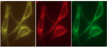
CYTO-ID® Green dye 能够很好的与RFP-LC3 蛋⽩兼容

A CYTO-ID® Green dye

B B:RFP-LC3

C CYTO-ID® Green dye与RFP-LC3结合

CYTO-ID Autophagy detection kit⾃噬分析试剂盒

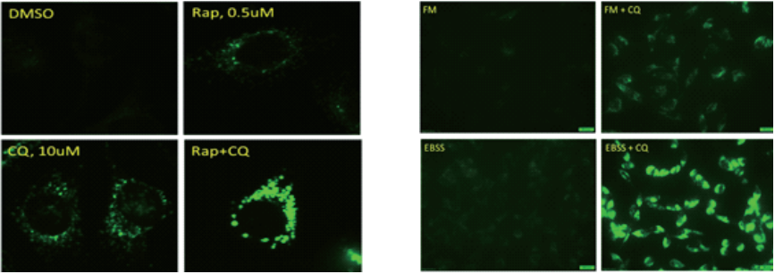
利⽤不同⽅法诱导的细胞⽤CYTO-ID Autophagy detection kit 荧光显微镜检测细胞⾃噬

CYTO-ID Autophagy detection kit⾃噬分析试剂盒

CYTO-ID Autophagy detection kit⽤于流式检测不同细胞⾃噬情况

产品信息:

货号 产品名称 规格
ENZ-51031 ENZ-51031 CYTO-ID Autophagy detection Kit 200 Flow cytometey assay

or 125microscopy assay

1*96 well

更多详情请咨询Enzo Life Sciences代理商-上海金畔生物

CYTO-ID® Autophagy detection kit 自噬检测试剂盒

CYTO-ID® Autophagy detection kit 自噬检测试剂盒

CYTO-ID® Autophagy detection kit     ENZ-51031-0050   ENZ-51031-K200

CYTO-ID自噬检测试剂盒

该试剂盒可在活细胞内定量分析细胞自噬,不需要转染。

适用于流式细胞术和显微镜。

可用于高通量筛选自噬的激活剂和抑制剂。

利用专利性的染料,可选择性的对前自噬体,自噬体和自噬性溶酶体进行染色。


文献引用

ABT-888 enhances cytotoxic effects of temozolomide independent of MGMT status in serum free cultured glioma cells: R. K. Balvers, et al.; J. Transl. Med. 13, 74 (2015), Application(s): Assay, Full Text

Bardoxolone methyl induces apoptosis and autophagy and inhibits epithelial-to-mesenchymal transition and stemness in esophageal squamous cancer cells: Y.Y. Wang, et al.; Drug Des. Devel. Ther. 9, 993 (2015), Application(s): Flow Cytometry,AbstractFull Text

Coffee and caffeine potentiate the antiamyloidogenic activity of melatonin via inhibition of Aβ oligomerization and modulation of the Tau-mediated pathway in N2a/APP cells: L.F. Zhang, et al.; Drug Des. Devel. Ther. 9, 241 (2015), Application(s): Flow Cytometry, AbstractFull Text

Evaluation of Antitumor Effects of Folate-Conjugated Methyl-β-cyclodextrin in Melanoma: K. Motoyama, et al.; Biol. Pharm. Bull. 38, 374 (2015), Application(s): Fluorescence Microscopy, AbstractFull Text

Induction of apoptosis and autophagy via sirtuin1- and PI3K/Akt/mTOR-mediated pathways by plumbagin in human prostate cancer cells: Z.W. Zhou, et al.; Drug Des. Devel. Ther. 9, 1511 (2015), Application(s): Assay, AbstractFull Text

Methicillin-Resistant Staphylococcus aureus Adaptation to Human Keratinocytes: G. Soong, et al.; MBio. 6, (2015),Application(s): Assay, AbstractFull Text

MiR-29b replacement inhibits proteasomes and disrupts aggresome+autophagosome formation to enhance the antimyeloma benefit of bortezomib: S. Jagannathan, et al.; Leukemia 29, 727 (2015), Application(s): Detection of autophagy by fluorescence microscopy in multiple myeloma cell lines, AbstractFull Text

Plumbagin induces G2/M arrest, apoptosis, and autophagy via p38 MAPK- and PI3K/Akt/mTOR-mediated pathways in human tongue squamous cell carcinoma cells: S. T. Pan, et al.; Drug Des. Devel. Ther. 9, 1601 (2015), Application(s): Assay,AbstractFull Text

Pro-apoptotic and pro-autophagic effects of the Aurora kinase A inhibitor alisertib (MLN8237) on human osteosarcoma U-2 OS and MG-63 cells through the activation of mitochondria-mediated pathway and inhibition of p38 MAPK/PI3K/Akt/mTOR signaling pathway: N. K. Niu, et al.; Drug Des. Devel. Ther. 9, 1555 (2015), Application(s): Assay, AbstractFull Text

The investigational Aurora kinase A inhibitor alisertib (MLN8237) induces cell cycle G2/M arrest, apoptosis, and autophagy via p38 MAPK and Akt/mTOR signaling pathways in human breast cancer cells: J. P. Li, et al.; Drug Des. Devel. Ther. 9, 1627 (2015), Application(s): Assay, AbstractFull Text

The pan-inhibitor of Aurora kinases danusertib induces apoptosis and autophagy and suppresses epithelial-to-mesenchymal transition in human breast cancer cells: J.P. Li, et al.; Drug Des. Devel. Ther. 9, 1027 (2015), Application(s): Assay, Abstract;Full Text

A Novel CXCR3-B Chemokine Receptor-induced Growth-inhibitory Signal in Cancer Cells Is Mediated through the Regulation of Bach-1 Protein and Nrf2 Protein Nuclear Translocation : M. Balan & S. Pal; J. Biol. Chem 289, 3126 (2014), Application(s):Monitor autophagy in MCF-7 and T47D breast cancer cells by flow cytometry and fluorescence microscopy, Abstract;

Adaptive responses to glucose restriction enhance cell survival, antioxidant capability, and autophagy of the protozoan parasiteTrichomonas vaginalis: K. Y. Huang, et al.; Biochim. Biophys. Acta. 1840, 53 (2014), Abstract;