EpiCypher新品促销—— CUTANA™ CUT&Tag Kit升级版(V 2)

EpiCypher新品促销—— CUTANA™ CUT&Tag Kit升级版(V 2)

EpiCypher新品促销—— CUTANA™ CUT&Tag Kit升级版(V 2)


EpiCypher CUTANA™ CUT&Tag Kit升级版(Version 2)来了!升级版各方面表现都优于V1版本,

助您持续生成高质量的CUT&Tag数据。新品推广期间凡订购EpiCypher CUTANA™ CUT&Tag Kit,可享额外20%折扣优惠!

前30名下单的客户,还有好礼相送(加油鸭挂件一个)


促销时间:即日起-2024.9.25

促销代码:CTK20


 促销产品清单 

供应商

产品货号

产品名称

产品规格

EpiCypher     

14-1102       

CUTANA™ CUT&Tag Kit with Primer Set 1      

48 Reactions      

EpiCypher

14-1103

CUTANA™ CUT&Tag Kit with Primer Set 2

48 Reactions

 六个常见问题带您了解CUT&Tag新变革 

EpiCypher新品促销—— CUTANA™ CUT&Tag Kit升级版(V 2)

从单细胞图谱分析、空间表观基因组学到 FFPE 样本分析,CUT&Tag正在成为研究热点1-7。 CUT&Tag也是EpiCypher的研发重点。多年来,EpiCypher的团队开发并优化了多种表观基因组图谱检测方法,包括CUT&Tag、CUT&RUN和ChIP-seq。EpiCypher团队充分利用专业知识研发的CUTANA™ CUT&Tag试剂盒,使这项令人难以置信的技术能够走进全球更多的研究领域和实验室。CUTANA™ CUT&Tag 试剂盒第2版现已上市,而且各方面都优于之前的版本。


从表面上看,CUT&Tag非常简单8,9。Protein A/G的融合体在抗体标记的染色质上栓了一种过度活跃的Tn5转座酶。激活的Tn5插入接头序列并切割DNA,这一过程被称为标签化。在EpiCypher的Direct-to-PCR方法中,使用识别接头序列的引物从反应混合物中直接扩增标记DNA,绕过文库制备,提高对低细胞数的敏感性。整个测定过程使用的是与磁珠结合的完整细胞核,并对在使用8联排管时的方法进行了优化,从而实现高通量工作流程和自动化解决方案。


为了简化实验过程,EpiCypher团队开发了CUTANA™ CUT&Tag试剂盒,其中包括从细胞到纯化的测序文库所需的所有试剂。请注意,CUT&Tag仅推荐用于组蛋白翻译后修饰(PTMs)的定位。 欲进一步了解如何使用CUT&Tag与其姊妹技术CUT&RUN,请您参阅这篇文章:http://www.jinpanbio.com/xwzx_2376.html。


尽管已取得了一些进展,但CUT&Tag仍然是一项具有挑战性的表观基因组检测技术。为了使其方法更加可靠,EpiCypher研发团队不断测试CUTANA™ CUT&Tag实验方案,研究不同的缓冲液成分,调整反应条件,并测试了多种细胞类型。CUTANA™ CUT&Tag试剂盒的第二版展现了EpiCypher最新的优化成果。


✍ 为什么科研学者会对CUT&Tag感兴趣?

科研人员之所以对CUT&Tag非常感兴趣,是因为它可以让科学家在不降低数据质量的情况下简化实验流程,这是ChIP不可能做到的。自从CUT&Tag出现以来,研发人员终于能够开发出更高通量的染色质图谱分析。与ChIP-seq相比,CUT&Tag可以使用更少的细胞,花费更少的时间,对更多的反应进行多重测序,并生成更高分辨率的数据。


✍ 为什么CUT&Tag比ChIP-seq和CUT&RUN快得多呢?

由于各种原因,CUT&Tag比其他染色质图谱分析技术更快。从样本处理的角度看,CUT&Tag无需再多花时间优化细胞裂解、交联或染色质碎裂。CUT&Tag使用完整的细胞核,可在15分钟内从新鲜或冷冻的细胞中获取。然后将细胞核与固体支撑物(ConA磁珠)结合,这样就能限制样品的损失,并可使用磁力架快速清洗。这一特点使该方法与8联排管兼容,进一步加快了实验速度。

从实验方法的角度看,直接插入测序接头无需进行标准的文库准备步骤,不仅节省了一天的工作时间,而且减少了样品损失。大多数CUT&Tag实验流程中需要纯化DNA,然后进行 PCR,而EpiCypher的Direct-to-PCR策略允许实验人员直接从CUT&Tag反应中扩增标记的DNA。因为所有的实验过程都是在8联排管中进行的,所以实验人员可以将接头引物和PCR混合物直接加入到CUT&Tag反应管中。对于时间紧张的项目,实验人员可以在CUT&Tag第2天结束时加载测序仪,第3天就可以开始处理数据。对于科研人员来讲,这样的周转时间非常宝贵。


✍ 与ChIP-seq相比,为什么在CUT&Tag中可以使用更少的细胞呢?

因为简化了实验流程,降低了背景信号影响,所以CUT&Tag需要的起始细胞数量较少。在 ChIP-seq中,需要交联和染色质超声处理或片段化来制备input的染色质。在免疫沉淀(IP)步骤中,抗体被添加到染色质片段化池中。理想情况下,抗体只与靶标结合,但免疫沉淀几乎总是能回收非靶标片段,并在测序数据中引入背景信号或测序假象。科学家通常会增加细胞数量以克服背景信号影响并提高数据质量,但这种解决方法会限制低数量细胞的应用。


CUT&Tag使用与磁珠结合的完整细胞核,不需要按照传统的IP步骤进行实验,从而减少了背景信号影响。EpiCypher的方法还绕过了ChIP和标准文库制备中的多个DNA纯化步骤,有助于减少样本的损失。总之,与ChIP-seq相比,CUT&Tag可以减少约10倍的材料,这是非常不可思议的。


✍ 科研学者在使用CUT&Tag实验操作流程时最常见的问题是什么呢?

研究者进行CUT&Tag实验时遇到的主要问题是产量低甚至没有产量,这可能是由多种变量影响的。产率低通常是由样品预处理不佳、实验过程中样品损失、ConA磁珠结合不上以及反应混合问题造成的。这些问题都有关联,因此解决起来比较复杂。


样品制备不良的表现是细胞裂解、细胞核制备中有碎片或ConA磁珠结合后上清液中存在未结合的细胞核。如果样本预处理出现上述情况,就可能无法进行标签化,进而导致产量低。


混合不充分也是产量低的一个常见原因。保持磁珠在溶液中对检测的成功至关重要:它有助于确保抗体和pAG-Tn5的均匀分布,并有助于高效的indexing PCR。然而,在实验方案中的第2天,ConA 磁珠浆会变得粘稠且难以重悬,尤其是在标记之后。虽然在处理材料时应当温和,但如果不能很好的混合CUT&Tag反应物,就会严重降低产量。 EpiCypher实验方案详细说明了何时以及如何混合样品,以便最终稳定地回收CUT&Tag生成的文库。


✍ 新版CUT&Tag试剂盒有什么亮点呢?

版本更新的重点是帮助使用者持续生成高质量的CUT&Tag数据。EpiCypher的团队详细讨论了实验流程的每个步骤。比如,为什么缓冲液要使用某种成分或pH值?对细胞核或细胞生理有何影响?这些问题帮助EpiCypher团队找到了可以改进的关键点。


pAG-Tn5的表征表明,酶本身并不是影响产量问题所在,且标记反应是高效的,但样品在标记后的实验步骤中损失了。为此,EpiCypher团队对样品处理、缓冲液成分、方案步骤进行了广泛的头对头比较研究,并对优秀的竞争产品进行了测试。


这些实验揭开了问题的神秘面纱。TAPS Buffer中的低盐浓度导致了渗透性变化和细胞核裂解。混合技术也尤为重要,它是造成样品损失的原因之一。例如,加入SDS Release Buffer后,样品变得粘稠,无法移液。在实验方案的其他部分,由于涡旋使材料粘在离心管边上,也会导致样品损失。


根据实验结果,EpiCypher团队去掉了标记后的低盐 TAPS Buffer洗涤,取而代之的是含有生理盐的Pre-Wash Buffer,以保持细胞核的完整性。EpiCypher团队还完善了实验流程中具体的重悬浮和混合方法,以帮助指导使用者进行最佳操作。EpiCypher内部测试了修改后的CUT&Tag实验方案,结果发现新手和有经验的使用者的实验成功率都有所提高,这说明了CUT&Tag改进后的实用性。


✍ 这些实验操作流程的变化适用于正在使用第1版CUTANA™ CUT&Tag Kit或DIY CUT&Tag的科研人员吗?

是的。所有实验流程的更改都与CUTANATM CUT&Tag Kit的第1版以及 DIY CUT&Tag流程(https://www.epicypher.com/resources/protocols/cutana-pag-tn5-resources/)兼容。

您可以使用现有的试剂盒组分来按照第2版的实验流程操作,而不需要任何新材料!


需要注意的主要区别是试剂盒中去掉了TAPS Wash Buffer。之前使用TAPS Wash Buffer进行标记后洗涤,现在使用等体积的Pre-Wash Buffer洗涤。EpiCypher在该试剂盒中为使用者提供了充足的Pre-Wash Buffer来完成这一步骤。


 总 结 

CUTANA™ CUT&Tag Kit版本的更新反映了EpiCypher严格的研发工作。EpiCypher团队将继续完善CUT&Tag和CUT&RUN的相关研究,包括针对新应用和研究领域的优化。如果您有其他疑问,第2版的实验操作流程(https://www.epicypher.com/resources/protocols/cutana-cut-and-tag-kit-manual/)也许能解决您的问题,也欢迎联系EpiCypher中国代理商上海金畔生物咨询。


 参考文献 

1. Janssens DH et al. Scalable single-cell profiling of chromatin modifications with sciCUT&Tag. Nat Protoc 19, 83-112 (2024). https://doi.org/10.1038/s41596-023-00905-9.


2. Bartosovic M et al. Single-cell CUT&Tag profiles histone modifications and transcription factors in complex tissues. Nat Biotechnol 39, 825-35 (2021). https://doi.org/10.1038/s41587-021-00869-9.


3. Wu SJ et al. Single-cell CUT&Tag analysis of chromatin modifications in differentiation and tumor progression. Nat Biotechnol 39, 819-24 (2021). https://www.doi.org/10.1038/s41587-021-00865-z.


4. Deng Y et al. Spatial-CUT&Tag: Spatially resolved chromatin modification profiling at the cellular level. Science 375, 681-6 (2022). https://doi.org/10.1126/science.abg7216.


5. Zhang D et al. Spatial epigenome-transcriptome co-profiling of mammalian tissues. Nature 616, 113-22 (2023). https://doi.org/10.1038/s41586-023-05795-1.


6. Henikoff S et al. Direct measurement of RNA Polymerase II hypertranscription in cancer FFPE samples. bioRxiv 2024.02.28.582647 (2024). https://doi.org/10.1101/2024.02.28.582647.


7. Henikoff S et al. Epigenomic analysis of formalin-fixed paraffin-embedded samples by CUT&Tag. Nat Commun 14, 5930 (2023). https://doi.org/10.1038/s41467-023-41666-z.


8. Kaya-Okur HS et al. CUT&Tag for efficient epigenomic profiling of small samples and single cells. Nat Commun 10, 1930 (2019). https://doi.org/10.1038/s41467-019-09982-5.


9. Kaya-Okur HS et al. Efficient low-cost chromatin profiling with CUT&Tag. Nat Protoc 15, 3264-83 (2020). https://doi.org/10.1038/s41596-020-0373-x.

如需了解更多详细信息或相关产品,

请联系EpiCypher中国代理商上海金畔生物 

请联系EpiCypher中国代理商-上海金畔生物 
EpiCypher——优质核小体的选择

EpiCypher——优质核小体的选择

越来越多的证据表明,核小体是体外表征许多染色质调节因子的最佳底物。随着携带完全确定组蛋白修饰的重组核小体的出现,为下一代染色质研究提供了新的或更好的方法(如染色质结合蛋白分析、酶分析、抗体谱分析)。EpiCypher拥有令人印象深刻的产品目录,其中包括83个独特的重组核小体库存,并且有能力生产定制设计核小体,质量高、周期短,助力您的研究!


在科研实验中,如果您要使用重组核小体进行染色质实验,则需考虑以下几点:


1、用于开发修饰组蛋白的方法。确保使用无疤痕方法整合所需的PTMs是很重要的,这种方法可以重现天然组蛋白结构。EpiCypher 使用几种不同的方法获得修饰的组蛋白,所有方法都会无疤痕整合组蛋白修饰。

为什么这很重要呢?许多市售的重组核小体是使用组蛋白PTM类似物构建的,如甲基赖氨酸类似物(MLAs) 10,其会导致修饰位点的氨基酸序列发生变化。这些非天然组蛋白修饰类似物已被证明会破坏与染色质调节蛋白和组蛋白PTM特异性抗体的相互作用,并且这对研究生理机制来讲是不理想的。因此,使用这些方法合成的核小体时应格外谨慎11-13。


2、修饰组蛋白的纯度。组装完全定义的同质核小体的下一步是对修饰组蛋白进行严格的质量控制。

在质控结果中,HPLC 迹线应显示是单一洗脱物质,表明组蛋白纯度>95%;平行高分辨率质谱(HRMS)应在预期质量的1道尔顿范围内显示一个单峰,没有任何意义的额外电荷质量(m/z)信号(例如图2A)。EpiCypher的所有修饰组蛋白均通过HPLC和HRMS分析进行验证。

为什么这很重要呢?不必要的物质(如甲硫氨酸氧化)可能引起结构改变,并影响静电相互作用或疏水相互作用,从而损害下游核小体组装的效率。


EpiCypher——优质核小体的选择


3、组装核小体的质量控制(QC)指标。DNA组装后核小体的质量验证对最终产品的信任保证至关重要。 

EpiCypher使用天然PAGE分析DNA上的dNuc组装,其中使用约150bp DNA的高效组装应该只产生单一物质,这相对于未组装的游离DNA,其迁移率降低(图2B,下图)。

为什么这很重要呢?因为被污染的游离DNA会诱导染色质修饰酶(如NSD2)的异常活性,所以必须避免。此外,次优组装可能导致样品的异质性混合,包括错误定位的核小体。

我们还使用考马斯染色对最终的dNucs进行SDS-PAGE分析,以确保四种组蛋白的化学计量相等(图2C,下图)。然后,我们通过免疫印迹证实了整合组蛋白PTM的存在(图2C,上图)。

为什么这很重要呢?如果其他种类的蛋白质污染或偏离1:1:1:1的比例,则可能表明组蛋白降解或组装不良,因此有必要重新解析每个组蛋白。而免疫印迹对于确定修饰是否存在于组蛋白的正确位置上非常重要。


核小体相关产品

分类

货号

产品名称

rNucs Human Recombinant 

Nucleosomes, No PTMs

16-0006

Mononucleosomes, biotinylated

16-0009

Mononucleosomes, non-biotinylated

16-0024

Mononucleosomes, desthiobiotinylated

16-0027

Tailless Nucleosomes, biotinylated

16-0023

Mononucleosomes, H3.1 ΔN2, biotinylated

16-0016

Mononucleosomes, H3.1 ΔN32, biotinylated

16-1016

Mononucleosomes, H3.1 ΔN32, non-biotinylated

16-0017

Mononucleosomes, H3.3 ΔN32, biotinylated

16-1017

Mononucleosomes, H3.3 ΔN32, non-biotinylated

16-0018

Mononucleosomes, H4 ΔN15, biotinylated

16-3004

Dinucleosomes, biotinylated

16-3104

Dinucleosomes, non-biotinylated

dNucs Designer Recombinant 

Nucleosomes with PTMs

16-0321

H3K4me1, biotinylated

16-0334

H3K4me2, biotinylated

16-1334

H3K4me2, non-biotinylated

16-0316

H3K4me3, biotinylated

16-1316

H3K4me3, non-biotinylated

16-0402

H3K4,K9me3, biotinylated

16-0403

H3K4,K27me3, biotinylated

16-0335

H3K4me3,K9,14,18ac, biotinylated

16-0325

H3K9me1, biotinylated

16-0324

H3K9me2, biotinylated

16-0315

H3K9me3, biotinylated

16-0338

H3K27me1, biotinylated

16-0339

H3K27me2, biotinylated

16-0317

H3K27me3, biotinylated

16-1317

H3K27me3, non-biotinylated

16-0397

H3.1K27me3,S28phos, biotinylated

16-0322

H3K36me1, biotinylated

16-0319

H3K36me2, biotinylated

16-0320

H3K36me3, biotinylated

16-1320

H3K36me3, non-biotinylated

16-0390

H3.3K36me3, biotinylated

16-0367

H3K79me1, biotinylated

16-0368

H3K79me2, biotinylated

16-0369

H3K79me3, biotinylated

16-0393

H4K12me1, biotinylated

16-0331

H4K20me1, biotinylated

16-0332

H4K20me2, biotinylated

16-0333

H4K20me3, biotinylated

16-1333

H4K20me3, non-biotinylated

vNucs Histone Variants

16-0013

H2AX, biotinylated

16-1013

H2AX, non-biotinylated

16-0366

H2AXS139phos, biotinylated

16-0014

H2AZ.1, biotinylated

16-1014

H2AZ.1, non-biotinylated

16-0015

H2AZ.2, biotinylated

16-0011

H3.3, biotinylated

16-0012

H3.3, non-biotinylated

Mutant Nucs Defined Amino 

Acid Substitutions

16-0029

H2AE61A, biotinylated

16-1029

H2AE61A, non-biotinylated

16-0030

H2AE92K, biotinylated

16-1030

H2AE92K, non-biotinylated

16-0031

H2BE105A,E113A, biotinylated

16-1031

H2BE105A,E113A, non-biotinylated

16-0349

Oncogenic Nucs (oncoNucs)

16-0350

H3.3K9M, biotinylated

16-1323

H3.3K27M, biotinylated

16-0323

H3.3K27M, non-biotinylated

16-0346

H3.3G34R, biotinylated

16-0347

H3.3G34V, biotinylated

16-0348

H3.3G34W, biotinylated

16-0344

H3.3K36M, biotinylated

Methyl DNA Nucs Nucleosomes 

with Methylated DNA

16-2043

Mononucleosomes, Recombinant, 

Hemi-methylated 199×601 DNA, biotinylated

16-2143

Mononucleosomes, Recombinant, 

Hemi-methylated 199×601 DNA, non-biotinylated

16-2044

Mononucleosomes, Recombinant, 

199×601 DNA, biotinylated

16-2144

Mononucleosomes, Recombinant, 

199×601 DNA, non-biotinylated

16-2045

Mononucleosomes, Recombinant, Symmetrically 

Methylated 199×601 DNA, biotinylated

EpiDyne® Chromatin Remodeling 

Assay Substrates

16-4201

EpiDyne FRET Nucleosome Remodeling 

Assay Substrate

16-4101

EpiDyne Nucleosome Remodeling Assay 

Substrate ST601-GATC1

16-4112

EpiDyne Nucleosome Remodeling Assay 

Substrate ST601-GATC1,2, biotinylated

16-4113

EpiDyne Nucleosome Remodeling Assay 

Substrate ST601-GATC1,2,3, biotinylated

16-4114

EpiDyne Nucleosome Remodeling Assay 

Substrate ST601-GATC1, 50-N-66, biotinylated

16-4115

EpiDyne Nucleosome Remodeling Assay 

Substrate ST601-GATC1,2, 50-N-66, biotinylated

16-4116

EpiDyne Nucleosome Remodeling Assay 

Substrate ST601-GATC1,2,3, 50-N-66, biotinylated

SNAP Spike-in Controls

19-1002

SNAP-CUTANA™ K-MetStat Panel

19-1001

SNAP-ChIP K-MetStat Panel

19-2001

SNAP-ChIP OncoStat Panel

19-3001

SNAP-ChIP K-AcylStat Panel

dCypher™ Nucleosome Panels

16-9001

dCypher™ Nucleosome Full Panel


想了解更多关于EpiCypher重组核小体技术和产品的信息吗?请联系金畔!


EpiCypher的注册商标和知识产权可见链接:https://www.epicypher.com/intellectual-property/。

本文中的所有其他商标和商品均为其各自公司所有。

本文翻译自链接:https://www.epicypher.com/resources/blog/finding-the-best-substrate-for-studying-histone-modifications/,如与原文有出入的地方,请以英文原文为准。

未经EpiCypher公司事先书面同意,本文件不得部分或全部复制。


关于EpiCypher公司:

EpiCypher是一家成立于2012年的表观遗传学公司。从专有组蛋白肽阵列平台EpiGold™开始,EpiCypher开发了一系列同类产品。同时,EpiCypher是重组核小体制造和开发的全球领导者。利用其独有技术,不断增加产品库中高纯度修饰重组核小体(dNucs™)产品。dNuc™多样性的产品为破译组蛋白编码和加速药物开发提供了强大的工具。

EpiCypher还将dNuc™技术广泛的应用于多种分析测定产品中,包括:SNAP-ChIP® Spike-in Controls(用于抗体分析和ChIP定量), EpiDyne® 底物(用于染色质重塑和抑制剂筛选及开发),dCyher™测定(用于探究表观遗传蛋白质-组蛋白PTM结合相互作用)。最近,EpiCypher还推出了针对ChIC、CUT&RUN和CUT&Tag的高灵敏度表观基因组图谱CUTANA™分析。


如需了解更多详细信息或相关产品,请联系EpiCypher中国代理商-上海金畔生物 
Epicypher热销产品——CUTANA™ ChIC/CUT&RUN Kit

Epicypher热销产品——CUTANA™ ChIC/CUT&RUN Kit

Epicypher热销产品——CUTANA™ ChIC/CUT&RUN Kit

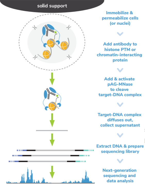
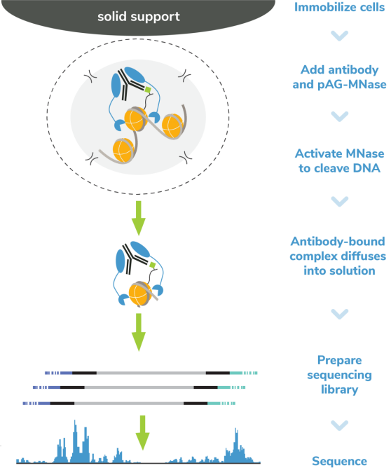
核酸酶靶向切割和释放 (CUT&RUN)技术是由Steven henikoff博士团队开发的一种染色质图谱分析方法,基于Ulrich Laemmli博士的染色质免疫切割技术 (ChIC),融合蛋白A与微球菌核酸酶 (pA-MNase),选择性原位切割与抗体结合的染色质。在CUT&RUN中,细胞或细胞核固定化在固相载体上,从溶液中分离出pAG-MNase裂解的DNA片段。该方法与二代测序(NGS)兼容,可提供高质量的组蛋白翻译后修饰(PTMs)和染色质相关蛋白(如转录因子;Figure 1)。

Epicypher热销产品——CUTANA™ ChIC/CUT&RUN Kit

ChIP-seq是组蛋白PTMs和染色质相关蛋白全基因组定位的主要方法。在这种方法中,染色质通过超声或酶消化破碎,然后免疫沉淀目标特异性片段。尽管进行优化,但ChIP-seq需要大量细胞(通常为105 – 106个细胞)而且需要深度测序input 染色质与免疫沉淀物质(通常为 >3000万 reads/次)来从背景中解析信号。

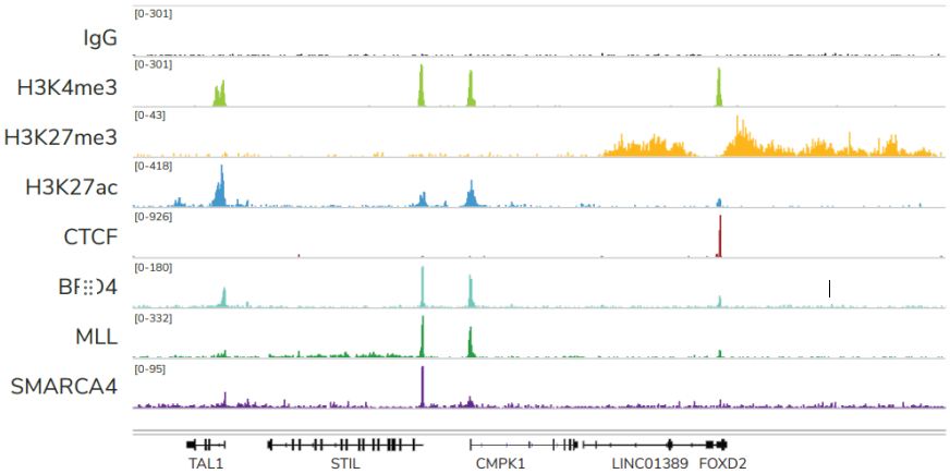
ChIC和CUT&RUN通过将基因组片段靶向释放到溶液中,彻底改变了染色质调控。通过这一创新,背景显著减少,允许使用少量细胞且每个反应仅需300 – 800万reads/次对组蛋白PTMs和染色质相关蛋白进行高分辨率基因组图谱的绘制(Figure 2)。简化的工作流程和节省的成本使ChIC/CUT&RUN适用于高通量研究表观遗传生物学。


Epicypher热销产品——CUTANA™ ChIC/CUT&RUN Kit

FIGURE 2 Representative genome browser tracks show CUTANA™ CUT&RUN results using 500,000 K562   cells.  Clear peaks with the expected distribution profile are observed using 3-8 million sequencing  reads per reaction for a variety of epigenetic targets, including histone PTMs (H3K4me3, H3K27me3,  H3K27ac), transcription factors (CTCF), epigenetic reader proteins (BRD4), writer enzymes (MLL1),   and chromatin remodelers (SMARCA4).  Rabbit IgG antibody shown as a negative control (top track).

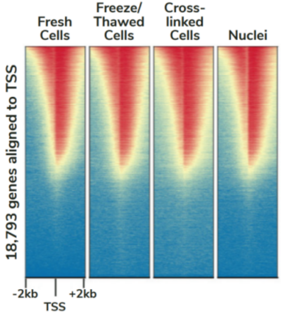
CUTANA™ChIC/CUT&RUN试剂盒包含48个反应的材料,专为多通道移液而设计,以实现CUT&RUN的高通量优势。该试剂盒包括阳性(H3K4me3)和阴性(Rabbit IgG)对照抗体,以及SNAP-CUTANA™ K-MetStat Panel (16个DNA条形码设计核小体携带广泛研究的赖氨酸甲基化PTMs)的分装分量。K-MetStat Panel加入到对照反应中,直接监测实验成功与否并帮助排除故障。此外,在pAG-MNase切割后,将剪切的E. coli DNA添加到所有反应中,以控制文库构建并使NGS标准化。该试剂盒与细胞和细胞核兼容,包括冻存和交联样品(Figure 3)。

Epicypher热销产品——CUTANA™ ChIC/CUT&RUN Kit

FIGURE 3 Heatmaps show CUT&RUN signal (red) and background (blue) of H3K4me3-enriched regions flanking annotated transcription start sites (TSS, +/- 2 kb). Gene rows are aligned across conditions, showing that genome-wide enrichment is preserved across sample types.


尽管建议从500,000个细胞开始,但只需使用5,000个细胞即可生成可比较的数据(Figure 4)。对照组的加入以及与不同靶类型、样本和低细胞数的兼容性,使该试剂盒成为染色质图谱实验的首选解决方案。

Epicypher热销产品——CUTANA™ ChIC/CUT&RUN Kit

FIGURE 4 Representative genome browser tracks for H3K4me3 (low abundance target) and H3K27me3  (high abundance target) CUT&RUN experiments using decreasing amounts of K562 cells. At 5,000   cells, data quality is largely indistinguishable from standard conditions (500,000 cells).

 保存条件 

OPEN KIT IMMEDIATELY and store components at room temperature, 4℃, and -20℃ as indicated (see User Manual corresponding to Kit Version 3). Stable for 6 months upon date of receipt.

Room Temperature (RT)

4℃

-20℃

8-strip Tubes

ConA Beads

5% Digitonin

0.5 M EDTA

E. coli Spike-in DNA

1 M Spermidine

100 mM Calcium Chloride

Bead Activation Buffer

SNAP-CUTANA™ K-MetStat Panel

SPRIselect Reagent

Manufactured by

Beckman Coulter Inc.

Pre-Wash Buffer

H3K4me3 Positive Control Antibody

0.1×TE Buffer

Stop Buffer

Rabbit IgG Negative Control Antibody

pAG-MNase

 数据示例 

Epicypher热销产品——CUTANA™ ChIC/CUT&RUN Kit

Figure 1: CUT&RUN DNA fragment size distribution analysis

CUT&RUN was performed as described in Figure 5. Library DNA was analyzed by Agilent Tapestation®. This analysis confirmed that mononucleosomes were predominantly enriched in CUT&RUN (~300 bp peaks represent 150 bp nucleosomes + sequencing adapters).

Epicypher热销产品——CUTANA™ ChIC/CUT&RUN Kit

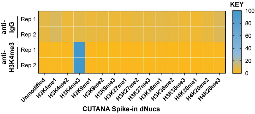
FIGURE 2 SNAP-CUTANA™ K-MetStat Spikein Controls

DNA-barcoded designer  nucleosomes (dNucs) representing 16 K-methyl PTMs: mono-, di-, and tri-methylation at H3K4, H3K9, H3K27, H3K36, and H4K20, as well as  unmodified control, were spiked into CUT&RUN  reactions prior to the addition of antibodies (IgG, H3K4me3). Spike-in barcodes were counted and  normalized from raw fastq files using the shell  script and analysis sheet available at  epicypher.com/19-1002. Barcodes for IgG (top;  normalized to total reads) and H3K4me3 (bottom; normalized to on-target) antibodies are  shown. The spike-ins confirmed optimal  experimental conditions (H3K4me3 antibody  specifically recovered the target dNuc, while IgG  showed no preferential enrichment).

Epicypher热销产品——CUTANA™ ChIC/CUT&RUN Kit

Figure 3: CUT&RUN genome-wide heatmaps

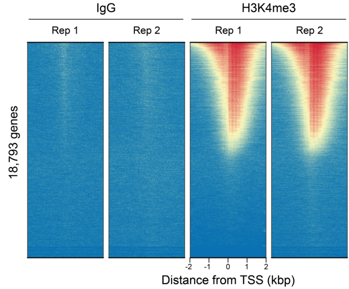
CUT&RUN was performed as described in Figure 5. Heatmaps show two replicates (“Rep”) of IgG (left) and H3K4me3 (right) kit control antibodies in aligned rows ranked by intensity (top to bottom) and colored such that red indicates high localized enrichment and blue denotes background signal.

Epicypher热销产品——CUTANA™ ChIC/CUT&RUN Kit

Figure 4: Representative gene browser tracks

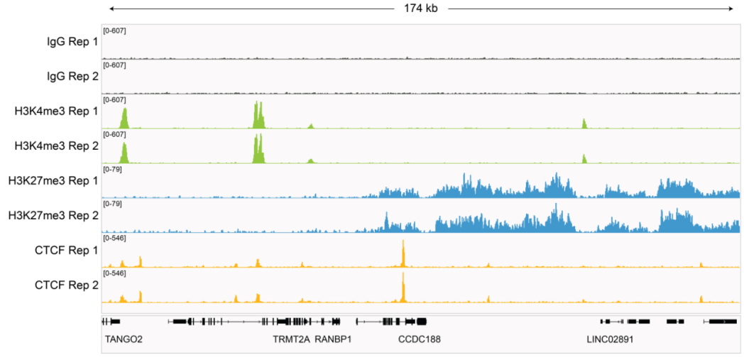
CUT&RUN was performed as described in Figure 5. A representative 174 kb window at the TRMT2A gene is shown for two replicates (“Rep”) of IgG and H3K4me3 kit control antibodies. Representative tracks are also shown for antibodies to H3K27me3 and the transcription factor CTCF. The CUT&RUN kit produced the expected genomic distribution for each target. Images were generated using the Integrative Genomics Viewer (IGV, Broad Institute).

Epicypher热销产品——CUTANA™ ChIC/CUT&RUN Kit

Figure 5: CUT&RUN methods

CUT&RUN was performed using the CUTANA™ ChIC/CUT&RUN Kit starting with 500k K562 cells with 0.5 µg of either IgG (EpiCypher 13-0042), H3K4me3 (EpiCypher 13-0041), H3K27me3 (EpiCypher 13-0055), or 0.125 µg of CTCF (EpiCypher 13-2014) antibodies in duplicate. Library preparation was performed with 5 ng of DNA (or the total amount recovered if less than 5 ng) using the CUTANA™ Library Prep Kit (EpiCypher 14-1001/14-1002). Libraries were run on an Illumina NextSeq2000 with paired-end sequencing (2×50 bp). Sample sequencing depth was 3.5 million reads (IgG Rep 1), 3.8 million reads (IgG Rep 2), 4.7 million reads (H3K4me3 Rep 1), 6.9 million reads (H3K4me3 Rep 2), 6.6 million reads (H3K27me3 Rep 1), 4.7 million reads (H3K27me3 Rep 2), 3.9 million reads (CTCF Rep 1) and 4.6 million reads (CTCF Rep 2). Data were aligned to the hg19 genome using Bowtie2. Data were filtered to remove duplicates, multi-aligned reads, and blacklist regions. 

 订购详情 

货号

产品名称

规格

14-1048

CUTANA™ ChIC/CUT&RUN Kit

48 Reactions


如需了解更多详细信息或相关产品,请联系EpiCypher中国代理商-上海金畔生物 
Epicypher新品 | 核酸提取缓冲液

Epicypher新品 | 核酸提取缓冲液

Epicypher新品 | 核酸提取缓冲液


CUTANA™ 细胞核提取缓冲液是CUT&RUN和CUT&Tag实验中,用于从培养的细胞和组织中提取细胞核的必需试剂。这种缓冲液可与多种真核细胞和组织兼容。以下是使用该缓冲液成功进行细胞核分离的细胞和组织类型:

mouse NIH3T3 fibroblast cells

human A549 non-small cell lung cancer (NSCLC) cells

human K562 leukemia cells

human NCI-H1299 non-small cell lung cancer (NSCLS cells)

human bone marrow derived macrophages

human TIG-1 fetal lung cells

human monocyte derived macrophages

human LoVo colorectal cancer cells

human MV-4-11 macrophage cells

human LNCaP prostate carcinoma cells

human SUM149 triple negative breast cancer (TNBC) cells

human renal primary cells

human GM24385 B-lymphocyte (aka HG002) cells

human peripheral blood mononuclear cells (PBMCs)

human MCF7 breast cancer cells

human intestinal tissue

human MDA-MB-231 breast cancer cells

human HEPM embryonic cells

human SK-MEL-2 melanoma cells


只需将试剂盒中的Pre-Nuclei Extraction Buffer和亚精胺与蛋白酶抑制剂混合即可完成该试剂的制备。CUTANA™ 细胞核提取缓冲液针对CUT&RUN和CUT&Tag实验流程进行了优化,从而获得高质量的细胞核,助力您的基因组图谱分析。


试剂盒组成

组分

货号

CUTANA™ Pre-Nuclei Extraction Buffer      

21-1026a     

1 M Spermidine

21-1026b


保存条件

将Pre-Nuclei Extraction Buffer 4ºC储存, 1 M亚精胺-20ºC储存。

自收到之日起可稳定储存6个月。


验证数据

Epicypher新品 | 核酸提取缓冲液

Figure 1: Nuclei Extraction

Nuclei were extracted from 3 different cell types: K562, NIH3T3, and LNCaP using the CUTANA™ Nuclei Extraction Protocol for CUT&RUN and CUT&Tag. Top 3 panels show cells before extraction. Starting cells are viable (bright white and round). Bottom 3 panels show nuclei extracted using the CUTANA™ Nuclei Extraction Buffer as indicated by positive Trypan Blue staining.


订购详情

货号

产品名称

规格

21-1026

CUTANA™ Nuclei Extraction Buffer

100 Reactions


如需了解更多详细信息或相关产品,请联系EpiCypher中国代理商-上海金畔生物 
Epicypher热销产品——Nucleosome, Recombinant Human, H3K4me3 dNuc, Biotinylated

Epicypher热销产品——Nucleosome, Recombinant Human, H3K4me3 dNuc, Biotinylated

EpiCypher是一家为表观遗传学和染色质生物学研究提供高质量试剂和工具的专业制造商。EpiCypher生产的重组单核小体 (H3K4me3) 由 147 个DNA碱基对缠绕在组蛋白的八聚体核心(H2A、H2B、H3.2 和 H4 各两个)上构成,是染色质的基本单位。Lowary和Widom[1]鉴定的147bp 601序列对组蛋白八聚体具有高亲和力,可用于核小体组装。H3K4me3 dNuc 在组蛋白H3.2的第4位含有三甲基赖氨酸。H3K4me3在110位处有一个Cys到Ala的取代。该DNA含有一个5’biotin-TEG基团。EpiCypher生产的重组单核小体适用于多种应用,包括用作酶分析、高通量筛选和抑制剂测试、染色质结合研究、蛋白质-蛋白质相互作用分析、结构研究和效应蛋白结合实验中的底物。

[1] Lowary and Widom J. Mol. Biol. (1998). PMID: 9514715

产品详情

宿主来源: Human

表达系统: E. coli & synthetic DNA

标签: Biotinylated

保存温度: Stable for six months at -80°C from date of receipt. For best results, aliquot and avoid freeze/thaws

产品形式: 0.977 mg/mL mononucleosome in 51.2 µL 10 mM Tris HCl pH 7.5, 25 mM NaCl, 1 mM EDTA, 2 mM DTT, 20% glycerol (27.3 µg protein, 50 µg DNA + protein)

验证数据

Epicypher热销产品——Nucleosome, Recombinant Human, H3K4me3 dNuc, Biotinylated

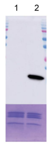
Figure 1: Western blot data

Western Analysis of H3K4me3 dNuc. Top Panel: Unmodified (EpiCypher 16-0006; 

Lane 1) and H3K4me3 (Lane 2) nucleosomes were probed with an anti-H3K4me3 

antibody and analyzed via ECL readout. Only the H3K4me3 sample produced a 

detectable signal. Bottom Panel: Detail from Coomassie stained gel showing unmodified 

(Lane 1) and H3K4me3 (Lane 2) nucleosomes.

Epicypher热销产品——Nucleosome, Recombinant Human, H3K4me3 dNuc, Biotinylated

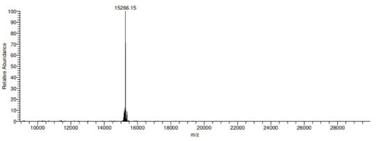
Figure 2: Mass spec data

Synthetic H3K4me3 histone analyzed by high resolution mass spectrometry. 

Expected mass = 15,267.77 Da. Determined mass = 15,266.15 Da.

Epicypher热销产品——Nucleosome, Recombinant Human, H3K4me3 dNuc, Biotinylated

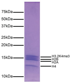
Figure 3: Protein gel data

Coomassie stained SDS-PAGE gel of proteins in H3K4me3 dNuc (1 µg) demonstrates 

the purity of histones in the preparation. Sizes of molecular weight markers and positions 

of the core histones (H2A, H2B, H3K4me3 and H4) are indicated.

Epicypher热销产品——Nucleosome, Recombinant Human, H3K4me3 dNuc, Biotinylated

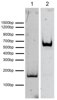
Figure 4: DNA gel data

H3K4me3 dNuc resolved via native PAGE gel and stained with ethidium bromide to 

visualize DNA. Lane 1: Free DNA (EpiCypher 18-0005; 100 ng). Lane 2: Intact 

H3K4me3 nucleosomes (400 ng).

订购详情

货号

产品名称

规格

16-0316

Nucleosome, Recombinant Human, H3K4me3 dNuc, Biotinylated

50 µg

16-0316-20

Nucleosome, Recombinant Human, H3K4me3 dNuc, Biotinylated

20 µg

如需了解更多详细信息或相关产品,请联系EpiCypher中国代理商-上海金畔生物 
Epicypher热销产品——SNAP-ChIP® K-MetStat Panel

Epicypher热销产品——SNAP-ChIP® K-MetStat Panel

由147个碱基对条形码widom601定位序列DNA缠绕在由大肠杆菌表达的重组人组蛋白组装而成的一组明显修饰的单核小体(组蛋白H2A、H2B、H3和H4各2个;accession numbers:H2A-P04908、H2B-O60814、H3.1-P68431或H3.2-Q71DI3*、H4-P62805),由1个未修饰的加上15个组蛋白H3或H4翻译后修饰(PTMs,通过专有的半合成方法创建)组成:H3K4、K9、K27和H4K20与me1、me2或me3。每个明显修饰的核小体都可以通过3’端的独特DNA序列(“barcode”)进行区分,该序列可以通过qPCR或二代测序进行破译。池中的16个核小体都被2种不同物种的DNA缠绕,每一种都含有独特的条形码(“A”和“B”,参考SNAP-ChIP手册)。适合用作ChIP、抗体特异性测试或效应蛋白结合实验的掺入质控品。

*组蛋白H3.2在110位包含一个Cys到Ala的替换。


产品详情

保存温度: Stable for six months at -20°C from date of receipt.

运输温度: DO NOT FREEZE!! Frozen cold packs.

产品形式: Purified recombinant mononucleosomes, containing a mixture of 16 (1 unmodified plus 15 unique) H3 and H4 PTMs in 10 mM sodium cacodylate pH 7.5, 100 mM NaCl, 1 mM EDTA, 50% glycerol (w/v), 1x Protease Inhibitor cocktail, 100 µg/mL BSA, 10 mM β-mercaptoethanol. Average molarity = 0.6 nM. MW = ~199382.1 Da (average MW of all 16 nucleosomes).


Epicypher热销产品——SNAP-ChIP® K-MetStat Panel

验证数据

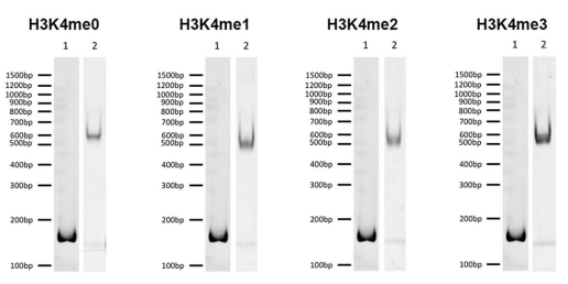
Fig 1. DNA Gel Data: Representative images for SNAP-ChIP K-MetStats (H3K4me0 = unmodified, H3K4me2, H3K4me3) run on a native PAGE gel and stained with ethidium bromide to visualize DNA. Lane 1: Free 147bp DNA used in nuclesome assembly (100 ng). Lane 2: Intact nucleosomes (200 ng) showing lack of free DNA. Identical experiments were performed for the entire K-MetStat Panel.

Epicypher热销产品——SNAP-ChIP® K-MetStat Panel

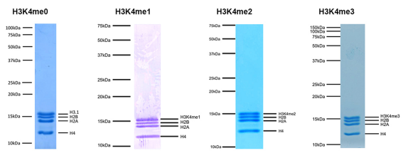
Fig 2. Protein Gel Data: Representative Coomassie stained PAGE gel for SNAP-ChIP K-MetStats (2 µg each of unmodified, H3K4me1, H3K4me2, H3K4me3) to demonstrate the purity of the histones in the preparation. Sizes of molecular weight markers and positions of the core histones (H2A, H2B, H3 and H4) are indicated. Identical experiments were performed for the remainder of the K-MetStat Panel.

Epicypher热销产品——SNAP-ChIP® K-MetStat Panel

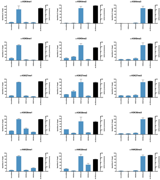
Fig 3. ChIP Data: Representative images for SNAP-ChIP K-MetStats (unmodified, H3K4me1, H3K4me2, H3K4me3) assayed in a chromatin immunoprecipitation (ChIP) experiment using commercially available ChIP grade antibodies (3 µg, n = 3). Quantitative Real-Time PCR (qPCR) for the DNA barcodes corresponding to unmodified (H3K4me0), H3K4me1, H3K4me2, and H3K4me3 nucleosomes show recovery of the barcodes corresponding to the expected antibody target. Identical experiments were performed for the remainder of the K-MetStat Panel (H3K9me1, H3K9me2, H3K9me3, H3K27me1, H3K27me2, H3K27me3, H3K36me1, H3K36me2, H3K36me3, H4K20me1, H4K20me2 and H4K20me3).

Epicypher热销产品——SNAP-ChIP® K-MetStat Panel

Fig 4. ChIP Data: Representative chromatin immunoprecipitation (ChIP) data using commercially available ChIP-grade antibodies targeting each PTM  in the K-MetStat panel.  The antibodies were assayed in a native ChIP experiment with 3 μg antibody added to 3 μg K-562 cell chromatin with  the K-MetStat Panel spiked-in prior to micrococcal nuclease digestion.  Quantitative real-time PCR (qPCR) was used to measure recovery of  duplicate DNA barcodes corresponding to the indicated panel nucleosomes (blue bars, x-axis).  The black bars map to the log scale on the right  y-axis and indicate the percentage of target immunoprecipitated relative to the input (a measure of the antibody efficiency).  In each case, the SNAP-ChIP spike-in confirmed that the antibodies recovered the expected histone PTM.  Enrichment of off-target PTMs is due to antibody  cross-reactivity.


订购详情

货号

产品名称

规格

19-1100       

SNAP-ChIP® K-MetStat Panel               

200 µL        

如需了解更多详细信息或相关产品,请联系EpiCypher中国代理商-上海金畔生物 

Epicypher热销产品——CUTANA™ E. coli Spike-in DNA

Epicypher热销产品——CUTANA™ E. coli Spike-in DNA

来源于Escherichia coli(E.coli)的片段DNA可以用作核酸酶靶向切割和释放(CUT&RUN)的实验标准化的spike-in对照。产品CUTANA™ E. coli Spike-in DNA含有足够的材料,可用于100-200个CUT&RUN样本(高丰度和低丰度靶标的范围)。

产品详情

保存温度: Stable for 2 years at -20°C from date of receipt. After resuspending, aliquots should be stored at -20°C.

运输温度: Room temp. Can go on frozen cold packs or dry ice.

产品形式: 100 ng lyophilized DNA. *NOTE: May not be visible.

验证数据

Epicypher热销产品——CUTANA™ E. coli Spike-in DNA

Figure 1: DNA gel data

E. coli fragment size distribution. Lane 1: gDNA extracted from JM101 E. 

coli cells (500 ng). Lane 2: Digested and purified CUTANA™ E. coli Spike-in 

DNA (500 ng) resolved via 2% E-Gel™ EX Agarose Gel. Migration positions 

of DNA molecular weight markers are indicated.

Epicypher热销产品——CUTANA™ E. coli Spike-in DNA

Figure 2: CUT&RUN sequencing data

CUTANA™ E. coli Spike-in DNA (1.0 ng) was added to CUT&RUN samples using 500,000 K562 cells enriched 

for a low abundance target (H3K4me3, EpiCypher 13-0041), a high abundance target (H3K27me3, EpiCypher 

13-0030) and an IgG negative control (EpiCypher 13-0042). Total numbers of paired-end sequencing reads, reads 

aligned to E. coli, and percentage of total sequencing reads aligned to E. coli spike-in DNA are shown. NOTE: 

Spike-in DNA amount should be optimized by the end user with the goal of E. coli DNA comprising ~1% (0.2 – 

5%) of total sequencing reads.

订购详情

货号

产品名称

规格

18-1401

CUTANA™ E. coli Spike-in DNA

100 ng

如需了解更多详细信息或相关产品,请联系EpiCypher中国代理商-上海金畔生物 
EpiCypher参与项目——SMARCAL1介导的肿瘤免疫逃避

EpiCypher参与项目——SMARCAL1介导的肿瘤免疫逃避

EpiCypher参与项目——SMARCAL1介导的肿瘤免疫逃避

资深作者Alberto Ciccia博士(左)和第一作者Giuseppe Leuzzi博士。


在新的博客系列中,我们将对话最近发表染色质研究工作的首席科学家们,了解他们的幕后工作。在EpiCypher,我们很感兴趣染色质技术是如何整合到不同领域的,如免疫学、细胞和基因治疗、发育生物学、代谢等。他们研究的主要结论是什么呢?相关论文对表观遗传学领域有何影响呢?


在本博客中,我们采访了来自哥伦比亚大学欧文医学中心遗传与发育系的Giuseppe Leuzzi博士和Alberto Ciccia副教授,他们最近在Cell上发表了题为 “SMARCAL1 is a dual regulator of innate immune signaling and PD-L1 expression that promotes tumor immune evasion”的文章。值得注意的是,这是一项高度合作的研究,来自世界各地很多个实验室都做出了贡献——包括EpiCypher。


Ciccia实验室研究了DNA损伤反应(DDR)在基因组完整性和癌症发展中的作用。在这项研究中,论文的第一作者Giuseppe Leuzzi使用CRISPR-Cas9开发了一种新的基因筛选方法,以评估DDR因子和染色质调节因子对先天免疫信号传导和PD-L1免疫检查点表达的影响。

利用这种方法,Leuzzi及其同事确定SMARCAL1是一种新的肿瘤免疫逃避调节因子,并且有希望成为更有效的免疫肿瘤学治疗靶点。


背景:免疫检查点阻断治疗与肿瘤耐药性

人体免疫系统旨在检测和破坏异常或感染的细胞,包括快速增殖的肿瘤细胞。许多细胞类型都参与这一过程。在本博客中,我们将重点关注靶向和杀死癌细胞的CD8+ 细胞毒性T细胞。


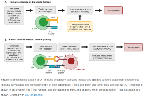
在许多癌症患者中,免疫系统被长期激活,导致CD8+ T细胞耗竭。耗竭的T细胞上调多种受体,如PD-1和CTLA-4受体,这些受体限制T细胞增殖、刺激和细胞毒性活性(图1A)。这些信号通路降低了CD8+ T细胞对抗癌症的效力,并被称为免疫检查点1-3。


靶向免疫检查点调节因子的药物代表了癌症免疫治疗的重大突破1。这些治疗方法,如PD-1抑制剂Keytruda(帕博利珠单抗),可阻断免疫检查点,并有助于恢复抗肿瘤免疫活性(图1A)。不幸的是,癌症经常会找到检查点通路或逃避免疫治疗(图1B)。了解肿瘤细胞如何调节这些信号对于开发改进的免疫疗法和生物标志物至关重要2。


EpiCypher参与项目——SMARCAL1介导的肿瘤免疫逃避


DNA损伤反应是如何影响免疫检查点治疗的呢?

基因组不稳定性在癌症发生、进展和治疗反应过程中发挥关键作用。最近的研究表明,化疗和放疗诱导的DNA损伤和DNA修复抑制会提高癌细胞中干扰素(IFN)的表达。这些先天免疫信号可以通过将T细胞募集到肿瘤微环境中来放大抗肿瘤免疫反应,最终导致肿瘤的排斥反应。因此,开发肿瘤内生性IFN治疗方法可能是癌症治疗的有效策略。


然而,延长的IFN信号传导会产生慢性炎症环境,从而导致T细胞耗竭和免疫抑制1-3。此外,IFN信号传导也可导致肿瘤中PD-L1过表达,从而使肿瘤细胞逃避免疫反应和检查点治疗(图1B)。


与作者问答

Leuzzi等人探索了调节肿瘤细胞中PD-L1和炎性细胞因子表达的新机制。如何利用IFN信号增强免疫治疗呢?他们最初的CRISPR筛选结果表明,SMARCAL1 DNA易位酶蛋白在这两个通路中均发挥重要作用。他们结合转基因细胞系、小鼠模型、生化和表观基因组学测定以及 TCGA 数据来研究SMARCAL1的功能。


在下面的对话中,Leuzzi和Ciccia在该领域的背景下讨论了他们的工作。为简洁明了,我们对内容进行了编辑。


Q:可以从您的研究文章中获得什么启示呢?

A:我们发现SMARCAL1是癌症免疫逃避和免疫治疗抵抗的主要调节因子。具体而言,SMARCAL1以肿瘤固有的方式发挥作用,并允许肿瘤细胞逃避内源性免疫系统和阻断免疫检查点的治疗。


Q:这是您要研究的内容吗?

A:该研究旨在发现新的癌症药物靶点,这些靶点可以增强人体的自然防御系统,同时还可以对抗免疫逃避机制,包括肿瘤细胞配体PD-L1的过度表达。人们认为,来自癌细胞先天免疫信号(如IFN)的上调伴随免疫抑制通路的下调,可恢复对免疫检查点阻断治疗的敏感性。


Q:您认为最令人惊讶的结果是什么呢?为什么?

A:我们最震惊的是SMARCAL1缺陷的影响,它诱导了癌症自主的IFN免疫信号传导,同时下调了PD-L1水平。该结果非常出乎意料,因为传统上认为IFN信号通路上调PD-L1是免疫检查点反应的一部分。这一观察结果违背了既定的预期,揭示了由SMARCAL1缺陷介导的不可预见的转折。


Q:这项工作是如何推动该领域发展的呢?

A:这项工作通过揭示之前未被识别的在肿瘤免疫逃避中起作用的SMARCAL1推动了这一领域的发展。值得注意的是,SMARCAL1使用独特的双重机制来调节IFN信号和PD-L1水平。首先,SMARCAL1通过限制内源性DNA损伤来抑制先天免疫信号传导。其次,SMARCAL1与JUN协同诱导PD-L1表达,使细胞逃避免疫系统的监视。这些发现为破坏逃避机制的靶向干预开辟了新途径,有可能提高癌症免疫治疗的有效性。


Q:CUT&RUN和EpiDyne试验为此提供了哪些重要的见解呢?

A:EpiCypher团队的专业知识,加上他们CUT&RUN chromatin mapping和EpiDyne™ nucleosome remodeling技术的使用,在揭示连接SMARCAL1和PD-L1表达的分子机制方面发挥了至关重要的作用。CUT&RUN实现了SMARCAL1结合位点的精确定位,揭示了其与JUN在转录活性染色质区域的合作。EpiDyne分析有助于评估SMARCAL1染色质的重塑能力。总之,这些技术为SMARCAL1如何影响染色质图谱和调节PD-L1免疫检查点基因提供了综合分子表征。


总结和结论

这篇激动人心的论文将DNA损伤反应和染色质调节与肿瘤免疫逃避和免疫治疗耐药性联系起来,从而为肿瘤免疫治疗的发展迎来了新的机遇。非常感谢Leuzzi 和Ciccia博士花时间与我们分享他们的工作,强烈推荐阅读他们的整篇论文(https://linkinghub.elsevier.com/retrieve/pii/S0092867424000102)。


参考文献

1. Pardoll DM. The blockade of immune checkpoints in cancer immunotherapy. Nat Rev Cancer 12, 252-64 (2012). https://doi.org/10.1038/nrc3239.

2. Fares CM et al. Mechanisms of Resistance to Immune Checkpoint Blockade: Why Does Checkpoint Inhibitor Immunotherapy Not Work for All Patients? Am Soc Clin Oncol Educ Book 39, 147-64 (2019). https://doi.org/10.1200/EDBK_240837.

3. Klapp V et al. The DNA Damage Response and Inflammation in Cancer. Cancer Discov 13, 1521-45 (2023). https://doi.org/10.1158/2159-8290.CD-22-1220

4. Leuzzi G et al. SMARCAL1 is a dual regulator of innate immune signaling and PD-L1 expression that promotes tumor immune evasion. Cell (2024). https://doi.org/10.1016/j.cell.2024.01.008


EpiCypher的注册商标和知识产权可见链接:https://www.epicypher.com/intellectual-property/。

本文中的所有其他商标和商品均为其各自公司所有。

本文翻译自链接https://www.epicypher.com/resources/blog/nuc-narratives-smarcal1,如与原文有出入的地方,请以英文原文为准。

未经EpiCypher公司事先书面同意,本文件不得部分或全部复制。


关于EpiCypher公司:

EpiCypher是一家成立于2012年的表观遗传学公司。从专有组蛋白肽阵列平台EpiGold™开始,EpiCypher开发了一系列同类产品。同时,EpiCypher是重组核小体制造和开发的全球领导者。利用其独有技术,不断增加产品库中高纯度修饰重组核小体(dNucs™)产品。dNuc™多样性的产品为破译组蛋白编码和加速药物开发提供了强大的工具。

EpiCypher还将dNuc™技术广泛的应用于多种分析测定产品中,包括:SNAP-ChIP®Spike-in Controls(用于抗体分析和ChIP定量), EpiDyne®底物(用于染色质重塑和抑制剂筛选及开发),dCyher™测定(用于探究表观遗传蛋白质-组蛋白PTM结合相互作用)。最近,EpiCypher还推出了针对ChIC、CUT&RUN和CUT&Tag的高灵敏度表观基因组图谱CUTANA™分析。


如需了解更多详细信息或相关产品,请联系EpiCypher中国代理商-上海金畔生物 

Epicypher热销产品——EpiDyne-FRET Nucleosome Remodeling Assay Substrate

Epicypher热销产品——EpiDyne-FRET Nucleosome Remodeling Assay Substrate

当与酶活性重塑复合物如SMARCA4、SMARCA2和ACF(如EpiCypher货号15-1014、15-1015、15-1013)配对时,该产品提供核小体重塑的高通量荧光读数。EpiDyne®-FRET单核小体由在大肠杆菌中表达的重组人组蛋白(组蛋白H2A-Cy5、H2B、H3.2和H4各两种;accession numbers::H2A-P4908;H2B-O60814;H3.2-Q71DI3;H4-P62805)组装而成,该组蛋白由212个DNA碱基对缠绕,并携带5’Cy3、Widom 601核小体定位元件和重塑时暴露的DpnII限制性内切酶基序(GATC)[1]。H2A在残基120位处具有Thr-Cys取代,其中Cy5是共轭的。H3.2在残基110位处具有Cys-to-Ala取代。

[1] Lowary & Widom J Mol. Biol. (1998) PMID: 9514715.


产品详情

保存温度:Stable for six (6) months at -80°C from date of receipt. For best results, aliquot and avoid multiple freeze/thaws.

运输温度:Dry ice.

产品形式:Purified recombinant mononucleosomes, (21.8 µg protein weight, 50 µg DNA+protein) in 44.2 µL of 10 mM Tris-HCl pH 7.5, 25 mM NaCl, 1 mM EDTA, 2 mM DTT, 20% glycerol. MW = 240,650 Da. Molarity = 4.7 µM.


验证数据

Epicypher热销产品——EpiDyne-FRET Nucleosome Remodeling Assay Substrate

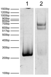
Figure 1: DNA Gel Data

EpiDyne-FRET substrate resolved via native PAGE gel and 

stained with ethidium bromide to visualize DNA. Lane 1: Free 

DNA (100 ng). Lane 2: Intact EpiDyne-FRET nucleosomes 

(400 ng).

Epicypher热销产品——EpiDyne-FRET Nucleosome Remodeling Assay Substrate

Figure 2: Protein Gel Data

Coomassie stained PAGE gel of proteins in EpiDyne-FRET 

substrate (1 µg) demonstrates the purity of histones in the 

preparation. Sizes of molecular weight markers and positions

of the core histones (H2A-Cy5, H2B, H3.2, and H4) are 

indicated.

Epicypher热销产品——EpiDyne-FRET Nucleosome Remodeling Assay Substrate

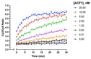
Figure 3: Nucleosome Remodeling Data

ACF1/ATP-dependent nucleosome remodeling reaction. EpiDyne-FRET 

nucleosomes (15 nM) were incubated with ACF1 chromatin remodeler

 (EpiCypher 15-1013; indicated concentrations) in the presence or absence 

of 1 mM ATP. Upon the addition of ATP, reactions were immediately read 

in an Envision Multilabelplate reader (PerkinElmer). Data are presented 

as the mean of the Cy3/Cy5 ratio (N=2).

订购详情

货号

产品名称

规格

16-4201   

EpiDyne-FRET Nucleosome Remodeling Assay Substrate   

50 µg   

如需了解更多详细信息或相关产品,请联系EpiCypher中国代理商-上海金畔生物 

Epicypher热销产品——Nucleosome, Recombinant Human, H2BK120ub1 dNuc, Biotinylated

Epicypher热销产品——Nucleosome, Recombinant Human, H2BK120ub1 dNuc, Biotinylated

EpiCypher是一家为表观遗传学和染色质生物学研究提供高质量试剂和工具的专业制造商。EpiCypher生产的在E. coli中表达的重组人单核小体(组蛋白H2A、H2B、H3和H4各2个;accession numbers:H2A-P04908;H2B-O60814;H3.1-P68431;H4-P62805)由601位处的147个DNA碱基对缠绕在组蛋白上构成。组蛋白H2B(由专门的半合成方法产生)在120位处含有泛素赖氨酸。核小体是染色质的基本单位,Lowary和Widom鉴定的601序列是一个含有147碱基对的序列,对组蛋白八聚体具有高亲和力,可用于核小体组装,并含有5’biotin-TEG基团。适合用作酶筛选分析或效应蛋白结合实验的底物。

[1] Lowary and Widom J. Mol. Biol. (1998). PMID: 9514715

产品详情

标签: Biotinylated

保存温度:Stable for six months at -80°C from date of receipt. For best results, aliquot and avoid multiple freeze/thaws.

产品形式: H2BK120ub dNuc (14.4 µg protein weight, 25 µg DNA + protein) in 22.5 µL 10 mM Tris HCl pH 7.5, 25 mM NaCl, 1 mM EDTA, 2 mM DTT, 20% glycerol. Molarity = 5.12 μM. MW = 216,985.9 Da.

验证数据

Epicypher热销产品——Nucleosome, Recombinant Human, H2BK120ub1 dNuc, Biotinylated

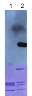
Figure 1: Western blot data

Western Analysis of Nucleosome, Recombinant Human, H2BK120ub. 

Top Panel: Unmodified H3 (Lane 1) and H2BK120ub containing nucleosomes 

(Lane 2) were probed with an anti-H2BK120ub antibody and analyzed via ECL

 readout. Only the H2BK120ub sample produced a detectable signal. Bottom 

Panel: Detail from Coomassie stained gel showing unmodified nucleosomes 

(Lane 1) and H2BK120ub nucleosomes (Lane 2).

Epicypher热销产品——Nucleosome, Recombinant Human, H2BK120ub1 dNuc, Biotinylated

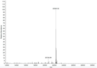
Figure 2: Mass spec data H2BK120ub protein analyzed by high 

resolution mass spectrometry. Expected mass = 22,321.8 Da. Determined 

mass = 22,320.3 Da.

Epicypher热销产品——Nucleosome, Recombinant Human, H2BK120ub1 dNuc, Biotinylated

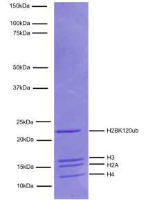
Figure 3: Protein gel data

Coomassie stained PAGE gel of proteins in Nucleosome, Recombinant 

Human, H2BK120ub (1 μg) to demonstrate the purity of the histones in the 

preparation. Sizes of molecular weight markers and positions of the core 

histones (H2A, H2BK120ub, H3.1, and H4) are indicated.

Epicypher热销产品——Nucleosome, Recombinant Human, H2BK120ub1 dNuc, Biotinylated

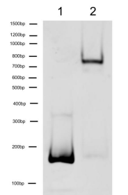
Figure 4: DNA gel data

Nucleosome, Recombinant Human, H2BK120ub dNucs resolved via native 

PAGE and stained with ethidium bromide to visualize DNA. Lane 1: Free 

DNA (100 ng). Lane 2: Intact H2BK120ub nucleosome (400 ng).

订购详情

货号

产品名称

规格

16-0396   

Nucleosome, Recombinant Human, H2BK120ub1 dNuc, Biotinylated   

25 µg   

如需了解更多详细信息或相关产品,请联系EpiCypher中国代理商-上海金畔生物