5-Aminosalicylic acid 5-氨基水杨酸

5-Aminosalicylic acid 5-氨基水杨酸

货号:
IA0250

品牌:
Jinpan
5-Aminosalicylic acid  5-氨基水杨酸

暂无详情
产品简介
MDL MFCD00007877
EC EINECS 201-919-1
别名 美沙拉嗪; ?氨水杨酸
CAS 89-57-6
分子式 C7H7NO3
分子量 153.14
纯度 HPLC≥98%
单位
生物活性 5-Aminosalicylic acid 是一种特异性的 PPARγ 激动剂,还抑制 p21-激活激酶1 (PAK1) 和 NF-κB。[1-3]
In Vitro 5-氨基水杨酸 (5-ASA) 是PPARγ的特异性激动剂, 只有PPARγ而非PPARα或PPARδ诱导p65降解。 5-氨基水杨酸诱导p65蛋白的降解, 表明PPARγ的E3泛素连接酶活性。 5-氨基水杨酸还在mRNA水平抑制PAK1, 这暗示了独立于PPARγ配体激活的另外机制。 5-氨基水杨酸通过抑制PAK1阻断肠上皮细胞 (IECs) 中的NF-κB[1]。用不同浓度 (10-1000μmol/ L) 的5-氨基水杨酸 (5-ASA) 或尼美舒利预处理12-96小时, 以剂量和时间依赖的方式抑制HT-29结肠癌细胞的生长。然而, 5-氨基水杨酸或尼美舒利的抑制没有统计学意义。当用不同剂量的5-氨基水杨酸和尼美舒利预处理时, HT-29结肠癌细胞的生长受到剂量依赖性的抑制。组合的5-氨基水杨酸 (终浓度100μM) 和尼美舒利 (终浓度10-1000μM) 以剂量依赖性方式抑制HT-29结肠癌细胞的增殖, 比相应剂量的尼美舒利更有效。类似地, 组合尼美舒利 (终浓度100μM) 和5-氨基水杨酸 (终浓度10-1000μM) 也剂量依赖性地抑制这些细胞的增殖, 比相应剂量的5-氨基水杨酸更有效[2]。
In Vivo 5-氨基水杨酸 (5-ASA) 在异种移植肿瘤模型中具有抗肿瘤作用。为了评价5-氨基水杨酸的体内抗肿瘤作用, 用50mM的5-氨基水杨酸每天处理植入HT-29结肠癌细胞的SCID小鼠连续21天。在治疗结束时, 与仅用GW9662治疗的对照小鼠或小鼠相比, 在接受5-氨基水杨酸的SCID小鼠中观察到肿瘤重量和体积减少80-86%。在5-氨基水杨酸处理10天后, 已经可检测到5-氨基水杨酸的抗肿瘤作用。用5mM的5-氨基水杨酸处理的小鼠获得了类似的结果。通过同时腹膜内施用GW9662, 在21天完全消除5-氨基水杨酸的抗肿瘤发生作用。因此, 观察到的5-氨基水杨酸的抗肿瘤作用至少部分依赖于PPARγ[3]。
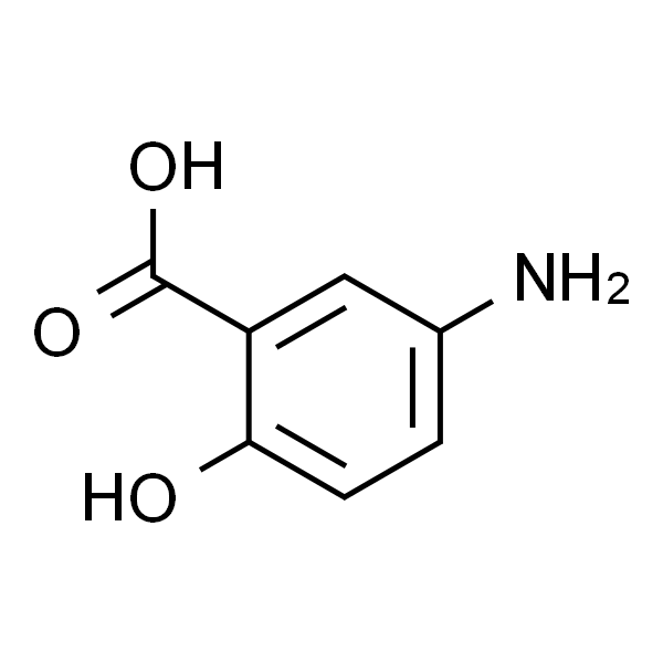
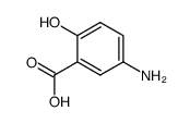
SMILES OC1=C(C(O)=O)C=C(N)C=C1
靶点 PAK1��PPAR��NF-��B
动物实验 小鼠[3]使用6至7周龄无病原体的BALB / c SCID小鼠。将用GW9662预处理或不处理24小时的人结肠癌细胞 (107 HT-29细胞) 皮下植入动物的侧腹。细胞接种后两天, 通过瘤周注射每天施用5-氨基水杨酸 (5或50mM) 处理小鼠10或21天。通过每日腹膜内注射GW9662 (1mg / kg /天) 评估PPARγ在5-氨基水杨酸处理期间的作用。对照组接受盐水而不是5-氨基水杨酸。每周检查小鼠三次以进行肿瘤发展。在10或21天杀死后, 计算肿瘤大小和体积。在石蜡包埋之前对肿瘤进行加权以进行组织学检查。
细胞实验 通过MTT测定法测量细胞抑制效应。用0.25%胰蛋白酶溶液分离HT-29结肠癌细胞5分钟。随后, 将细胞接种到96孔板 (1×10⁶细胞/孔) 上, 补充10%FCS并使其附着24小时, 然后加入试验化合物 (5-氨基水杨酸10, 50, 100, 500) , 和1000μM; 尼美舒利; 和它们的组合) 。将测试化合物在无血清培养基中稀释。然后将细胞在培养基或不同浓度的药物中孵育48小时, 加入20μL在PBS中的MTT溶液 (5g / L) 。 4小时后, 除去各孔中的培养基, 加入120μL0.04mM盐酸异丙醇, 稍微浓缩10分钟。用ELISA读数器在490nm处测量染料吸收。每个浓度使用5个孔或作为对照组。另一方面, 将细胞接种到96孔板 (1×10 6个细胞/孔) 上并使其附着24小时, 然后用试验化合物 (5-氨基水杨酸, 尼美舒利及它们的组合) 处理。最终浓度为100μM。将相同的培养基加入对照组中, 然后测量染料摄取。每个测试化合物或对照组使用5个孔[2]。
数据来源文献 [1]. Dammann K, et al. PAK1 modulates a PPARγ/NF-κB cascade in intestinal inflammation. Biochim Biophys Acta. 2015 Oct; 1853 (10 Pt A) :2349-60.

[2]. Fang HM, et al. 5-aminosalicylic acid in combination with Nimesulide inhibits proliferation of colon carcinoma cells in vitro. World J Gastroenterol. 2007 May 28; 13 (20) :2872-7.

[3]. Rousseaux C, et al. The 5-aminosalicylic acid antineoplastic effect in the intestine is mediated by PPARγ. Carcinogenesis. 2013 Nov; 34 (11) :2580-6.

备注 以上数据均来自公开文献, Jinpan暂未进行独立验证, 仅供参考。These protocols are for reference only. Jinpan does not independently validate these methods.
规格 50mg 10mM*1mL (in DMSO) 500mg

是一种特异性的 PPARγ 激动剂,还抑制 p21-激活激酶1 (PAK1) 和 NF-κB。

5-Aminosalicylic Acid

5-Aminosalicylic AcidCAS号: 89-57-6分子式: C7H7NO3分子量: 153.14描述纯度储存/保存方法别名外观可溶性/溶解性靶点In vitro(体外研究)In vivo(体内研究)

产品描述
描述

5-Aminosalicylic acid 是一种特异性的 PPARγ 激动剂,还抑制 p21-激活激酶1 (PAK1) 和 NF-κB。
纯度
>98%
储存/保存方法
Store at -20℃ for one year(Powder);Store at 2-4℃ for two weeks;Store at -20℃ for six months after dissolution.
基本信息
别名
5-氨基水杨酸;美沙拉嗪;;Mesalamine;5-ASA;Mesalazine
外观
White to gray solid
可溶性/溶解性
DMSO : ≥ 6 mg/mL (39.18 mM)
生物活性
靶点
IKK
In vitro(体外研究)
Mesalamine inhibits the enzyme 3-hydroxysteroid dehydrogenase, involved in the reversible conversion between DHP and THP, and therefore may affect the local actions of DHP and THP in the brain. Mesalamine, an anti-inflammatory aminosalicylate, dose-dependently inhibits IL-1-stimulated NF-kappaB-dependent transcription without preventing IkappaB degradation or nuclear translocation and DNA binding of the transcriptionally active NF-kappaB proteins, RelA, c-Rel, or RelB. Mesalamine is found to inhibit IL-1-stimulated RelA phosphorylation. Mesalamine increases cell adhesion which is measured by cell adhesion assay and transcellular-resistance measurement. Mesalamine treatment restores membranous expression of adhesion molecules E-cadherin and β-catenin. Mesalamine or sulfasalazine (2 mM), but not sulfapyridine, significantly reduces the expression of the TC22 transcript and significantly reduces the expression of TC22 protein in a dose-dependent and reversible manner. Mesalamine induces membranous expression of E-cadherin and increases intercellular adhesion. Mesalamine activity modulates E-cadherin glycosylation and increases both mRNA and protein levels of GnT-III and its activity as detected by increased E4-lectin reactivity. Mesalamine (0.1-1 mM) shows considerable inhibition of peroxynitrite-mediated luminol chemiluminescence in a dose-dependent fashion, suggesting that Mesalamine is able to directly scavenge the peroxynitrite. Mesalamine only at higher concentration (1 mM) inhibits the hydroxyl radical adduct while shifting Electron paramagnetic resonance (EPR) spectra.
In vivo(体内研究)
5-Aminosalicylic acid (5-ASA) has an antineoplastic effect in a xenograft tumor model. To evaluate the in vivo antineoplasic effect of 5-Aminosalicylic acid, SCID mice engrafted with HT-29 colon cancer cells are treated daily for 21 consecutive days with 5-Aminosalicylic acid at 50 mM. At the end of the treatment, a reduction of 80-86% of tumor weight and volume is observed in SCID mice receiving 5-Aminosalicylic acid compared with control mice or mice treated with GW9662 alone. The antineoplastic effect of 5-Aminosalicylic acid is already detectable after 10 days of 5-Aminosalicylic acid treatment. Similar results are obtained with mice treated with 5-Aminosalicylic acid at 5 mM. Antitumorigenic effect of 5-Aminosalicylic acid is completely abolished at 21 days by simultaneous intraperitoneal administration of GW9662. Thus, the observed antineoplastic effect of 5-Aminosalicylic acid is at least partially dependent on PPARγ.

分子结构图

5-Aminosalicylic Acid