BioXCell最新文献引用

BioXCell最新文献引用

Nature Communications最新发布的文献 ”Contrast-enhanced ultrasound with sub-micron sized contrast agents detects insulitis in mouse models of type1 diabetes” 使用了BioXCellInVivoPlus anti-mouse CD4(Clone: GK1.5)抗体, 揭示了使用亚微米级造影剂的超声造影检测I型糖尿病小鼠模型中的胰岛素炎。

 BioXCell最新文献引用

摘要:In type1 diabetes (T1D) autoreactive T-cells infiltrate the islets of Langerhans, depleting insulin-secreting β-cells (insulitis). Insulitis arises during an asymptomatic phase, prior to clinical diagnosis of T1D. Methods to diagnose insulitis and β-cell mass changes during this asymptomatic phase are limited, precluding early therapeutic intervention. During T1D the islet microvasculature increases permeability, allowing nanoparticles to access the microenvironment. Contrast enhanced ultrasound (CEUS) uses shell-stabilized gas bubbles to provide acoustic backscatter in vasculature. Here, we report that sub-micron sized ‘nanobubble’ ultrasound contrast agents can be used to measure increased islet microvasculature permeability and indicate asymptomatic T1D. Through CEUS and histological analysis, pre-clinical models of T1D show accumulation of nanobubbles specifically within pancreatic islets, correlating with insulitis. Importantly, accumulation is detected early in disease progression and decreases with successful therapeutic intervention. Thus, sub-micron sized nanobubble ultrasound contrast agents provide a predicative marker for disease progression and therapeutic reversal early in asymptomatic T1D.


文章链接:https://www.nature.com/articles/s41467-020-15957-8


订购详情

产品名称

货号

规格

InVivoMAb anti-mouse CD4

BE0003-1

1/5/25/50/100mg

InVivoPlus anti-mouse CD4

BP0003-1

1/5/25/50/100mg


详情请咨询 BioXcell 中国代理上海金畔生物科技

全国服务热线: 15221999938            邮箱: sales@jinpanbio.com

深圳: 021-50837765      北京: 021-50837765      上海: 021-50837765          

广州:18024516375        香港: 021-50837765

代理品牌网站: www.jinpanbio.com

自主品牌网站: www.jinpanbio.cnet


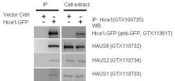
GFP抗体,实验应用广,100多篇文献引用

GFP抗体,实验应用广,100多篇文献引用

GFP抗体,实验应用广,100多篇文献引用 GFP抗体,实验应用广,100多篇文献引用

产品名称:GFP抗体

产品货号:GTX113617

宿主:Rabbit

克隆:Polyclonal

同种型:IgG

实验应用:WB, ICC/IF, IHC-P, IHC-Fr, IHC-Wm, IP, Dot, ELISA, IHC

产品属性

形式:Liquid

存储溶液:1XPBS (pH7), 1% BSA, 20% Glycerol

存放说明:Store as concentrated solution. Centrifuge briefly prior to opening vial. For short-term storage (1-2 weeks), store at 4ºC. For long-term storage, aliquot and store at -20ºC or below. Avoid multiple freeze-thaw cycles.

浓度:0.17 mg/ml (Please refer to the vial label for the specific concentration.)

免疫原:Full length GFP recombinant protein

纯化方式:Purified by antigen-affinity chromatography.

偶联:Unconjugated

RRID:AB_1950371

别名:Green Fluorescent Protein , GFP , eGFP , enhanced green fluorescent protein

注意事项:仅供实验室使用。不适用于人类或动物的任何临床,治疗或诊断用途。不适合动物或人类食用。

订购详情

品牌 产品名称 产品货号 规格
Genetex GFP antibody GTX113617 100μl
Genetex GFP antibody GTX113617-S 25μl

部分文献引用

Shu-Jung Chang et al. Elife 2022; 11 Typhoid toxin sorting and exocytic transport from Salmonella Typhi-infected cells.

实验应用 : WB

Guofu Zhu et al. Br J Pharmacol 2022; 179 (8) : 1716-1731 A novel peptide inhibitor of Dll4-Notch1 signalling and its pro-angiogenic functions.

实验应用 : IP

Gonzalo P Solis et al. Nat Commun 2022; 13 (1) : 2072 Local and substrate-specific S-palmitoylation determines subcellular localization of Gαo.

实验应用 : WB

Mikkel ? N?rg?rd et al. Sci Rep 2022; 12 (1) : 496 A new transgene mouse model using an extravesicular EGFP tag enables affinity isolation of cell-specific extracellular vesicles.

实验应用 : WB

Miete C et al. Nat Commun 2022; 13 (1) : 674 Gαi2-induced conductin/axin2 condensates inhibit Wnt/β-catenin signaling and suppress cancer growth.

实验应用 : WB

更多详细信息,请咨询GeneTex代理商-上海金畔生物

InvivoGen带您读文献-自噬与天然免疫

InvivoGen带您读文献-自噬与天然免疫

自噬与其作用

自噬(Autophagy)是胞捕获废物,去除物和物循的三大主要机制之一,另外两种分是蛋白酶降解(proteasomal degradation)和吞噬(phagocytosis)。在自噬过程中,胞浆中的小分子被新形成的自噬体包住,随后在一种特殊的溶酶体中消化,代谢产物将重新被释放到胞浆中。

自噬,常常被称作巨噬,可将胞质转化成养物,保障胞在饥饿,应激等状态下维持大分子合成以及能量平衡。

此外,胞利用自噬调节特殊的信号蛋白活性,阻止损伤细胞器或长寿聚集倾向蛋白的积累;去除细胞内病原的威胁。因此,自噬已成为天然免疫的重要部分。

InvivoGen 提供自噬研究相关产品

自噬诱导剂:Metformin,Rapamycin,MG-132, SAHA

自噬抑制剂:SP600125,U0126,Bafilomycin A1,SB202190等

自噬相关调控基因:ATG,LC3,BECN1,RAB7,TFEB等

自噬体相关细胞:HeLa-Difluo™ hLC3 CellsRAW-Difluo™ mLC3 Cells


InvivoGen带您读文献-自噬与天然免疫

HeLa-DiFluoTM cells were treated with 25 μM rapamycin alone or with 25 μM rapamycin and 500 nM bafilomycin A1 (to inhibit autophagosome/lysosome fusion). After 24 hour incubation, the cells were fixed with 2% PFA and analyzed by confocal microscopy. Note that both yellow (autophagosome) and red (autolysosome) puncta increase in panel viii, whereas most puncta in panel ix are yellow (autophagosome).


自噬信号通路

典的自噬信号通路通一系列特定的步骤进行。

InvivoGen带您读文献-自噬与天然免疫


InvivoGen带您读文献-自噬与天然免疫

自噬从分离膜的成核开始,逐将要清除的包成一个口袋。口袋的膜封后可形成一个双膜构小泡,被称自噬体(autophagosome),随后其外膜与溶酶体融合形成自噬溶酶体(autolysosome)。重新整合的内膜将捕获的胞内物质暴露于溶酶体水解酶,后者将材料分解成代谢产物组分[1,2]。

典的自噬通路需要一些列化保守基因的协调动作。小泡成核依赖于由Beclin1,Vps34和其它蛋白形成的III型磷脂酰肌醇-3羟基激酶(PI(3)K)复合体。

Atg7参与两条泛素化样结合通路:Atg5与Atg12合,将LC3化成磷脂乙醇胺PE)结合LC3-II形态。Atg5-Atg12与Atg15L1蛋白形成一个大复合体。两个结合系统都是形成自噬体的必要条件。

然而,我需要些或其它自噬蛋白也可多非自噬功能。因此,研究者最大的挑是从自噬蛋白的信号中区分出“真正”的自噬信号。


选择性自噬

饥饿或某些激状下,胞利用自噬消化和循内大的,非特异性组分。然而,自噬也参与免疫信号通路的调节(例如,被循活化蛋白限制胞因子的生),以及阻断炎症反(例如,在损伤的线粒体释放活性氧族(ROS)之前将其去除)。

了分离和去除特异性威胞利用选择性自噬构,在其中,被中的靶物首先被泛素标记。泛素化的物质通过LC3被拖入并与分离膜绑定,在众多捕获小体样蛋白(SLRs)中(如p62, optineurin,parkin或PINK1),根据捕获物质的不同,由其中一种蛋白参与捕获过程。


自噬与天然免疫

自噬与天然免疫信号通路有广泛的系-例如,损伤相关分子模式(DAMPs)和病原相关分子模式(PAMPs)的反应。研究表明,自噬与多种模式识别受体(PRRs)是调节与被调节的关系,这些受体包括Toll-like receptors (TLRs)Nod-like receptors (NLRs)RIG-I-like receptors (RLRs) cytosolic DNA sensors (CDS)  Stimulator of Interferon Genes (STING) 以及炎症小体。


InvivoGen带您读文献-自噬与天然免疫

这种协作包括正向和负向调节机制,以确保阻止急性炎症反应的产生过度炎症。有趣的是,自噬与其信号蛋白与炎症紊乱(如克罗恩氏综合症),一些癌症和自身免疫病(如硬化症和系统性红斑狼疮)有关[3]。另外,自噬缺陷与神经疾病有关,例如帕金森病症是由蛋白结晶累积造成。


自噬TLRs

TLRs由细胞膜(TLR1, TLR2,TLR4,TLR5 和 TLR6) 和小胞体 TLR3, TLR7, TLR8, TLR9 和 TLR13)受体组成,每个TLR都可对自身,病原或肿瘤的同源DAMPs和PAMPs做出应答。代表性的TLR配体包括脂多糖(LPS TLR4),鞭毛蛋白(TLR5)和 肽葡聚糖(TLR2)等细菌复合物;DNA(TLR9)和RNA(TLR3,TLR7,TLR8和TLR13)等核酸。活化TLRs可诱导产生促炎性细胞因子,在某些情况下,则诱导产生1型干扰素(IFNs)。

TLR诱导的自噬依赖于适配体蛋白MyD88和TRIF;两条信号通路均与Beclin-1直接作用[6]。在巨噬细胞等免疫细胞中,TLR配体刺激诱导吞噬体和自溶酶体的形成[7],并且,在细菌和病毒感染小鼠模型过程中,体内实验(in vivo)证明TLR诱导自噬形成[8]。

自噬调节系统可将DNA和RNA投放到含TLRs的小胞体中。事实上,有报道证明在树突细胞中,自噬和与之密切相关的过程LC3相关吞噬作用(LAP)TLR7和TLR9识别核酸过程起到重要作[7]

有趣的是,TLR诱导的自噬与一些疾病密切相关。例如,TLR3和TLR4诱导的自噬与肺癌细胞的转移和入侵有关[9]。


自噬与NODs

有报道指出其它天然免疫受体与自噬相呼应,不过有可能是细胞类别特异性现象[10,11]。这些受体包括细胞浆受体,如核苷酸结合寡聚结构域蛋白1和2(NOD1 和 NOD2),后者可分别识别肽聚糖衍生物D-glutamyl-meso-diaminopimelic acid (iE-DAP)和胞壁酰二肽(MDP)。在巨噬细胞中,NOD1和NOD2与Atg16L1协作,诱导自噬;在树突细胞中,细菌配体活化NOD2可导致生成吞噬体[7]。


自噬和cGAS/STING 通路

cGAS/STING 通路是天然免疫细胞浆中识别内源和外源DNA的主要通路。cGAS感受器识别dsDNA(和DNA/RNA混合体),而后产生信使分子2’,3’-cGAMP,一种可活化适配蛋白STING的环二核苷酸,而后,STING驱动1型干扰素和促炎性因子的产生。入侵的微生物也可通过释放的环二核苷酸(CDNs)直接活化STING。

cGAS/STING通路与自噬息息相关。例如,在结合分歧杆菌(M. tuberculosis)感染后,cGAS/STING可激活自噬和1型干扰素产生[7],并且参与此病原的选择性自噬(Selective autophagy)[12]

有趣的是,有报道证明,cGAS识别胞浆DNA后,可由p62依赖的选择性自噬降解[13]。另外,在M. bovis感染过程中,胞浆DNA感受器AIM2可抑制STING诱导的自噬[14]

有些报道指出,在某些情况下,cGAS和STING分别与自噬蛋白互动,而这些相互之间的影响并没有被完全解析。例如,cGAS与Beclin-1的相互作用可终止2’,3’-cGAMP的产生[15],因此阻止STING的活化和1型干扰素的过表达。此外,STING的转运似乎牵扯Atg9a[16]


自噬与RLR

天然免疫的兵工厂中,还有一样武器叫RIG-I样受体 (RLRs),可识别胞浆中自身或外源RNA,包括病毒RNA。主要的RLRs是RIG-I,识别短双链RNA;和MDA5,识别长双链RNA。活化后,这两个受体都要通过激活适配蛋白MAVS诱导产生1型干扰素和促炎性因子。

虽然RLRs与自噬相互影响的报道很少,但是有迹象表明,在细胞质中,自噬负向调节RLRs应答内源或外源RNA,从而限制1型干扰素的产生。例如,自噬蛋白Atg5和Atg12的结合可干扰双链RNA感受器(MDA5  RIG-I)和适配器蛋白MAVS的信号传递[17]。与之相似的是,泛素特异性蛋白酶19(USP19)(被证明可正向调节自噬),可通过Beclin-1依赖的方式抑制RIG-IMAVS的相互作用,从而削弱干扰素信号通路[17]


自噬,炎症小体和线粒体

炎症小体是一个复合体,由一个天然免疫感受器(AIM2NLRP1NLRP3NLRC4)加上一个适配蛋白,凋亡相关斑点样蛋白CARD(ASC),和pro-Caspase1。每个炎症小体根据其装配的感受器命名。

炎症小体对DAMPs和PAMPs比如胞浆DNA(AIM2),MDP(NLRP1),尿酸(NLRP3)和鞭毛蛋白(NLRC4)作出应答,从而促使促炎性反应。活化炎症小体诱导炎性白介素1和18的前体产生

研究证实,自噬可调节活化的炎症小体,从而限制过度炎症反应。自噬可通过消化炎症小体介导的白介素前体(如,pro-IL-1)和循环的炎症小体组分(如,NLRP3,AIM2和ASC)来直接限制炎症小体通路[18]

另外,自噬可通过间接的方式阻止炎症小体活化,前者降解损伤的线粒体,从而阻止炎症小体活化配体-线粒体DNA(mtDNA)和活性氧族(ROS)的释放。最近研究指出,在受损的线粒体膜上,活化的MAVS可直接与LC3相互作用,诱导产生自噬体,从而去除有害细胞器[19]。因此,细胞缺失自噬蛋白Atg5呈现受损细胞器堆积现象,结果导致1型干扰素加剧产生[20]


结论

2016年诺贝尔生理或医学奖,颁发给发现自噬机制的大隅良典,以表示对这一细胞代谢过程在健康和疾病上至关重要作用的极大肯定。

然而,自噬与天然免疫信号通路相互作用的研究,才刚刚开始。


参考文献

1. Levine B. & Kroemer G., 2008. Autophagy in the pathogenesis of disease. Cell. 132(1):27-42.

2. Mizushima N. et al., 2008. Autophagy fights disease through cellular self-digestion. Nature. 451(7182):1069-75.

3. Netea-Maier et al., 2016, Modulation of inflammation by autophagy: Consequences for human disease. Autophagy. 12(2): 245–260.

4. Xu Y. et al., 2007. Toll-like receptor 4 is a sensor for autophagy associated with innate immunity. Immunity. 27(1):135-44.

5. Delgado MA. et al., 2008. Toll-like receptors control autophagy. EMBO J. 27(7):1110-21.

6. Shi CS. & Kehrl JH., 2008. MyD88 and Trif target Beclin 1 to trigger autophagy in macrophages. J Biol Chem. 283(48):33175-33182.

7. Shibutani et al., 2015. Autophagy and autophagy-related proteins in the immune system. Nat Immunol. 16(10):1014:1024.

8. Zhan et al., 2014, Autophagy facilitates TLR4- and TLR3-triggered migration and invasion of lung cancer cells through the promotion of TRAF6 ubiquitination. Autophagy. 10(2):257-68.

9. Zhan et al., 2014. Autophagy facilitates TLR4- and TLR3-triggered migration and invasion of lung cancer cells through the promotion of TRAF6 ubiquitination. Autophagy. 10(2):257-268.

10. Kroemer G. et al., 2010. Autophagy and the integrated stress response. Mol Cell. 40:280–293.

11. Deretic V., 2012. Autophagy as an innate immunity paradigm: expanding the scope and repertoire of pattern recognition receptors. Curr. Opin. Immunol. 24(1):21-31.

12. Watson et al., 2015. The Cytosolic Sensor cGAS Detects Mycobacterium tuberculosis DNA to Induce Type I Interferons and Activate Autophagy. Cell Host & Microbe. 17:1-9.

13. Chen et al., 2016. TRIM14 Inhibits cGAS Degradation Mediated by Selective Autophagy Receptor p62 to Promote Innate Immune Responses. Mol Cell. 64:1-15.

14. Liu et al., 2016. AIM2 inhibits autophagy and IFN-β production during M. bovis infection. Oncotarget. 7(30):46972-46987.

15. Liang et al., 2014. Crosstalk between the cGAS DNA Sensor and Beclin-1 Autophagy Protein Shapes Innate Antimicrobial Immune Responses. Cell Host & Microbe. 15:228-238.

16. Saitoh et al., 2009. Atg9a controls dsDNA-driven dynamic translocation of STING and the innate immune response. PNAS. 106(49):20842–20846.

17. Jin et al., 2016. USP19 modulates autophagy and antiviral immune responses by deubiquitinating Beclin-1. EMBO J. 35(8): 866–880.

18. Harris et al., 2017. Autophagy and Inflammasomes. Mol Immunol. In press: http://dx.doi.org/10.1016/j.molimm.2017.02.013

19. Sun et al., 2016. MAVS maintains mitochondrial homeostasis via autophagy. Cell Disc. 2:16024.

20. Chan & Gack, 2015. RIG-I-like receptor regulation in virus infection and immunity. Curr. Opin. Virol. 12:7-14.


订阅金畔微信公众号随时掌握第一手新消息促销 产品试用 奖学金 都信手拈来,还不快扫一扫我们的公众号,或直接搜索关注“上海金畔生物”,更多惊喜等你发现!


上海金畔生物科技有限公司

InvivoGen带您读文献之“STING与病毒防御”

InvivoGen带您读文献之“STING与病毒防御”

天然免疫是宿主的第一道防线。站在“长城”上的天然免疫感受器实时监控并识别外来入侵的病原微生物,并启动抗微生物免疫应答。当微生物进入宿主细胞,活化不同的胞浆感受器,而由不同的胞浆感受器启动的信号最终都被STING受体接收,后者诱导产生I型干扰素和促炎性细胞因子。随着STING抗病毒应答研究的深入,更多的疑问也被提出,例如STING是如何与不同胞浆核酸感受器或适配器协同完成抗病毒免疫应答InvivoGen公司提供全系列STING相关产品,协助您深入开展STING研究。


InvivoGen提供的STING相关产品

STING 配体: 2’3’-cGAMP – 3’3’-cGAMP – c-di-GMP – c-di-AMP …

STING 变异株: STING-WT – hSTING-HAQ – hSTING-H232 …

STING 敲除细胞:RAW-Lucia ISG-KO-STING Cells – THP1-Dual KO-STING Cells…

STING 报告基因细胞系:RAW-Lucia ISG Cells – THP1-Blue ISG Cells …

ELISA 试剂盒:LumiKine™ Xpress hIFN-α – LumiKine™ Xpress hIFN-β

更多产品信息,请访问www.invivogen.cn


STING 与病毒防御

宿主细胞接触病毒后会立即启动天然免疫应答,包括诱导产生I型干扰素和促炎性细胞因子,以便清除入侵的病原。最近研究发现,在病毒感染后,可被细胞内受体IFI16,cGAS和STING识别并激活I型干扰素信号通路,这样的防御机制涵盖DNA病毒和RNA病毒。研究HSV-1和CMV等DNA病毒时发现,病毒基因组DNA可以结合DNA感受器-IFI16和cGAS,从而揭示了外源基因组的DNA识别机制[1,13]研究证明病毒感染宿主细胞后,将其基因组DNA被释放到细胞内,后者可激活STING-TBK1依赖的免疫应答[2,3,4]也有研究指出,病毒感染后可以引起细胞应激,触发线粒体释放DNA到胞浆,活化cGAS-STING信号通路[22]。随着研究的深入与拓展,科研人员发现STING在RNA病毒应答中也扮演重要角色。过表达STING可以抑制新城疫病毒和禽流感病毒的复制,而这个防御机制并不依赖于RIG-I和cGAS[5,6]敲底STING的表达水平,也可以提高登革热病毒的复制[7,8]。丙型肝炎病毒调节STING的应答机制虽然存在一些小的争议,不过可以确定的是,丙型肝炎病毒刺激STING敲除的细胞并不能引起I型干扰素的产生[9,10]RNA病毒也存在一些含有DNA的核酸结构,逆转录病毒在复制过程中产生的反转录DNA中间体如RNA:DNA,单链DNA,亦激活DNA感受器。HIV-1在侵染细胞中释放的单链DNA可被cGAS识别,激活I型干扰素[11, 23]。在病毒感染过程中,不同来源的DNA活化cGAS-STING通路的机制还需要进一步探明。    

越来越多的迹象表明,被感染的细胞不仅可以识别DNA完成自身防御,同时将cGAMP通过缝隙连接(gap junction)传递到临近细胞,从而激活STING依赖的I型干扰素表达[24]另外,有报道指出病毒颗粒可装配cGAMP,从而传递并激活天然免疫应答[25]。以上研究揭示了STING在病毒防御中的核心作用,为预防或治疗传染性疾病提供新线索。


STING 与病毒拮抗免疫防御策略

在病毒和免疫系统共同进化的压力下,病毒学会了如何与免疫系统对抗并成功入侵宿主的技能。抑制I型干扰素应答是病毒入侵的关键机制。据报道,疱疹病毒家族通过不同的策略抑制STING-TBK1信号通路,如降解或锁住IFI16[4,13];抑制cGAS的酶活性[15];或抑制STING和TBK1的相互作用[16]。其它病毒也有对应STING通路的抑制策略。乙型肝炎病毒可以促使STING降解从而抑制其功能[17]DNA肿瘤病毒中,乳头瘤病毒的E7蛋白和腺病毒的E1A蛋白可拮抗cGAS-STING通路,从而抑制免疫应答[19]DNA病毒外,RNA病毒也具有调剂STING的能力。虽然这看起来违背常理,不过已发表的数据表明RNA病毒具有不同的抑制STING应答方式。登革热病毒编码的NS2B3蛋白酶可作用于人源STING的氨基酸末端,裂解人源STING,而鼠源STING对NS2B3蛋白酶具有抗性 [7,8]丙型肝炎病毒编码的NS4B蛋白可阻断STING和TBK1的相互作用[10]A型流感病毒的血凝素蛋白可阻止STING二聚体形成[6]; HIV干扰STING与TBK1的相互作用[21]。随着科研工作的深入,病毒逃避免疫识别的机制也逐渐清晰,研究重点需转向关于体内(in vivo)的感染机制。随着对致病性病毒疾病的认知不断积累,病毒感染细胞的作用靶点以及病毒逃避免疫防御的机制都是未来抗病毒新药研发的参考依据。


1. Horan KA, et al. 2013.Proteasomal degradation of herpes simplex virus capsids in macrophages releases DNA to the cytosol for recognition by DNA sensors. J Immunol.

2. Lam E1, et al. 2014. Adenovirus detection by the cGAS/STING/TBK1 DNA sensing cascade. J Virol.

3. Victor R. DeFilippis, et al. 2010.Human Cytomegalovirus Induces the Interferon Response via the DNA Sensor ZBP1. J Virol.

4. Li T, et al. 2013. Human cytomegalovirus tegument protein pUL83 inhibits IFI16-mediated DNA sensing for immune evasion. Cell Host Microbe.

5. Yuqiang Cheng, et al. 2015.Chicken STING Mediates Activation of the IFN Gene Independently of the RIG-I Gene. J Immunol.

6. Christian K. Holm. et al. 2016.Influenza A virus targets a cGAS-independent STING pathway that controls enveloped RNA viruses. Nat commun.

7. Chia-Yi Yu, et al. 2012. Dengue Virus Targets the Adaptor Protein MITA to Subvert Host Innate Immunity. PLoS Path.

8. Sebastian Aguirre, et al. 2012.DENV Inhibits Type I IFN Production in Infected Cells by Cleaving Human STING. PLoS Path.

9. Nitta S, et al. 2013. Hepatitis C virus NS4B protein targets STING and abrogates RIG-I-mediated type-I interferon-dependent innate immunity. Hepatology.

10. Qiang Ding, et. al. 2013. Hepatitis C virus NS4B blocks the interaction of STING and TBK1 to evade host innate immunity. J Hepatol.

11. Daxing Gao, et. al. 2013.Cyclic GMP-AMP Synthase Is an Innate Immune Sensor of HIV and Other Retroviruses. Science.

12. Sunnie M. Yoh, et. al. 2015.PQBP1 Is a Proximal Sensor of the cGAS-Dependent Innate Response to HIV-1. Cell.

13. Orzalli MH, et al. 2012.  Nuclear IFI16 induction of IRF-3 signaling during herpesviral infection and degradation of IFI16 by the viral ICP0 protein. PNAS.

14. Maria H Christensen, et al. 2016. HSV-1 ICP27 targets the TBK1-activated STING signalsome to inhibit virus-induced type I IFN expression. The EMBO Journal.

15. Wu JJ, et al. 2015. Inhibition of cGAS DNA sensing by a herpesvirus virion protein. Cell Host Microbe.

16. Ma Z, et al. Modulation of the cGAS-STING DNA sensing pathway by gammaher- pesviruses. PNAS 2015.

17. LiuY, et al. 2015. Hepatitis B virus polymerase disrupts K63-linked ubiquitination of STING to block innate cytosolic DNA-sensing pathways. J Virol.

18. McLaughlin-Drubin ME, Munger K. 2009. The human papillomavirus E7 oncoprotein. Virology.

19. Laura L, et al. 2015.  DNA tumor virus oncogenes antagonize the cGAS-STING DNA-sensing pathway. Science.

20. Xing Y, et al. 2013. The papain-like protease of porcine epidemic diarrhea virus negatively regulates type I interferon pathway by acting as a viral deubiquitinase. J Gen Virol.

21. Haitao Guo, et al. 2016. NLRX1 Sequesters STING to Negatively Regulate the Interferon Response, Thereby Facilitating the Replication of HIV-1 and DNA Viruses. Cell Host & Microbe.

22. West AP, et al. 2015.  Mitochondrial DNA stress primes the antiviral innate immune response. Nature.

23. Herzner AM, et al. 2015. Sequence-specific activation of the DNA sensor cGAS by Y-form DNA structures as found in primary HIV-1 cDNA. Nat Immunol.

24. Ablasser A, et al. 2013. Cell intrinsic immunity spreads to bystander cells via the intercellular transfer of cGAMP. Nature.

25. Gentili M, et al. 2015. Transmission of innate immune signaling by packaging of cGAMP in viral particles. Science.

订阅金畔微信公众号随时掌握第一手新消息促销 产品试用 奖学金 都信手拈来,还不快扫一扫我们的公众号,或直接搜索关注“上海金畔生物”,更多惊喜等你发现!

上海金畔生物科技有限公司