胶原蛋白诱导小鼠关节炎实验方案(二)

胶原蛋白诱导小鼠关节炎实验方案(二)

E. Collagen

胶原

因为去糖基化的胶原会影响关节炎的产生(11),同时一些未去除的杂质比如:胃蛋白酶可能会导致T 细胞刺激试验中产生假阳性(12),因此二型胶原应按规定高度纯化。如果按恰当的方法冻干的胶原-20℃避光保存的条件下,是非常稳定的。纯化的胶原溶于0.05M 浓度的醋酸中,终浓度为2-4mg/ml,温和搅拌,4℃过夜。胶原溶液可在4℃保存一周,此后需在-20℃保存。

Chondrex 公司提供较完整的不同免疫级别二型胶原. 比如,DBA/1小鼠对鸡或者牛的二型胶原蛋白有较强的免疫反应,然后C57B/6 小鼠只对鸡的二型胶原蛋白有免疫反应。

Catalog # Description
20011 Chick type II collagen,10mg

鸡II 型胶原蛋白

20012 Chick type II collagen, 5 ml x 2 mg/ml

鸡II 型胶原蛋白

20021 Bovine type II collagen, 10 mg

牛II 型胶原蛋白

20022 Bovine type II collagen, 5 ml x 2 mg/ml

牛II 型胶原蛋白

20031 Porcine type II collagen, 10mg

猪II 型胶原蛋白

20032 Porcine type II collagen, 5 ml x 2 mg/ml

猪II 型胶原蛋白

A. Preparing the Emulsion

乳剂单位制备

乳剂的质量对于诱发关节炎至关重要。乳剂的制备有多种方法。但是不推荐双注射器或超声波处理的方法。这些方法将导致乳剂不能够有效的、稳定的诱发关节炎。此外,超声处理方法,至少将胶原分成 2 个片段,这些小片段在正常体温下,很容易变性。

推荐使用电动的高速搅拌器:

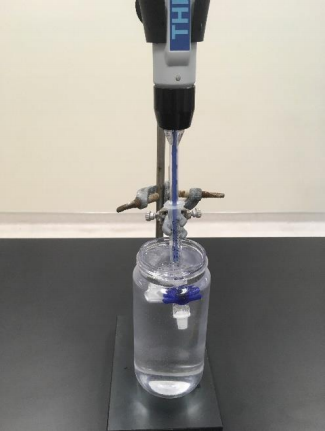
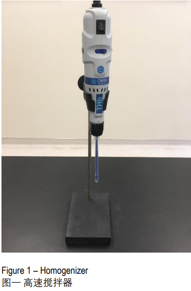
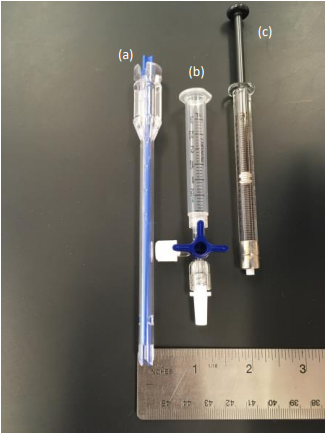
1.用高速搅拌器 (图一)(带直径≤5mm 的刀片,图二a)搅拌完全佐剂CFA (或用于增强剂注射的不完全佐剂(IFA))和胶原溶液 将注射器尖端用三通阀封闭 (图二b)。把注射器夹紧,固定在铁架台上,并冰浴 (图三)。最后一步很重要,因在搅拌过程中会发热,会引起蛋白变性,变性的蛋白不能够诱发关节炎(CIA)。

胶原蛋白诱导小鼠关节炎实验方案(二)

(图一 高速搅拌器)

胶原蛋白诱导小鼠关节炎实验方案(二)

(图二 (a) 搅拌器刀片-直径0.5cm, (b)带三通阀的注射器,(c)玻璃注射器)

胶原蛋白诱导小鼠关节炎实验方案(二)

(图三 带三通阀的注射器固定在支架上,冰浴)

2.加 1 体积(最多不超过注射器的25%)完全佐剂(不完全佐剂在加强免疫时用到)到注射器,再缓慢加入等体积的胶原溶液(0.05M 醋酸溶解的的胶原溶液,终浓度为 2mg/ml),边低速搅拌(1000-3000rpm)边滴入胶原溶液。

注:为保证高质量的免疫乳剂,一次量乳剂体积应不得超过注射器体积的50%。如果需要多量生产,可重复以上步骤。

3.继续高速混合(10,000 – 30,000rpm)2分钟。冷却5分钟,再重复高速混合和冷却步骤2-3次直到产生稳定的乳液。对于大体积乳剂制作(2-5ml)为了是乳液混合均匀,建议在混匀过程中上下移动搅拌刀片。

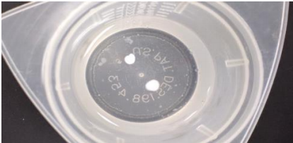
4.用针头替换三通阀,在装有冰水的烧杯里滴一滴乳剂,检测乳剂的稳定性。如果乳剂是稳定的,如图所示(图四),在水中是成团的,不散开。如果乳剂在水中散开,表明乳剂不稳定,再加几滴佐剂,重新混合再次进行测试。

胶原蛋白诱导小鼠关节炎实验方案(二)

(Figure 4 – 图四 完整稳定的乳剂漂浮在水面)

5.将乳剂转移到1ml的玻璃注射器 (图二C).不建议采用塑料注射器,因为很难做到一个准确的注射剂量。

注:1.通过用力甩注射器将乳剂中的气泡赶出注射器(活塞部向下)。否则很难做到一个精确的注射剂量。

        2.建议在乳剂制备好的一小时内完成注射。乳剂在使用之前存放在4°C。

B. Injection Site

免疫注射部位

在鼠尾根部皮下注射 0.1ml乳剂(100μg胶原每只老鼠(图五)。例如:从尾根部 2cm 处插入针头(25号或者27号针头,长度5/8英尺,针插入皮下,针头沿斜向上与鼠尾方向平行插入,直到针尖插入位置距尾根部0.5cm。每次注射前充分擦拭针头,防止乳液的露出。

如果要加强免疫,注射部位在尾根部 3cm 处,针尖插入皮下距鼠尾根部1.5cm处。加强免疫的注射部位应避开初始注射部位)。

注: Chondrex公司不建议使用背部皮下注射注射或腹腔注射胶原乳剂和佐剂,因为乳剂会引起腹腔和胸腔严重的炎症反应。

(图五 皮下注射乳剂)

如需完整资料或咨询相关问题,请联系Chondrex代理商–上海金畔生物

胶原抗体诱导小鼠关节炎模型之CAIA实验方案

胶原抗体诱导小鼠关节炎模型之CAIA实验方案

PROTOCOL TO INDUCE CAIA

CAIA实验方案

Chondrex公司建议尾静脉注射单克隆抗体合剂。对于易感品系,可采用腹腔注射,但是通过腹腔注射CAIA诱发的炎症严重程度往往较低,关节炎持续时间也较短。请根据实验目的选择合适的实验方案。

Catalog Number Amount
53100 100mg
53040 40mg
53010


A. Inducing CAIA without LPS

胶原抗体诱导的关节炎

在易感品系DBA/1和Balb/c小鼠中采用CAIA建立关节炎模型,根据动物供应商,动物周龄和体积大小的不同,每只小鼠需注射6-10mg单克隆抗体合剂。例如,初次注射免疫5mg抗体合剂后24小时再次注射5mg抗体合剂。关节炎会在二次免疫后的24-48小时后发生。研究员需要根据实验目的调整实验方案。

B. Inducing CAIA with LPS 

胶原抗体联用LPS诱导的关节炎

细菌毒素LPS(B细胞分裂素), MAM(支原体产生的T细胞分裂素)或SEB(金黄色葡萄球菌产生的T 细胞分裂素)和胶原抗体在诱发关节炎中具有协同效应。在这些毒素的存在下,诱导关节炎的抗体合剂的量可降低。例如,每只小鼠初次免疫注射1.5mg抗体合剂后第三天注射25-50µgLPS,对于易感品系小鼠(Balb/c, DBA/1,B10.RIII, and C.B-17 scid/scid mice),关节炎的发生率高达100%。在C57BL/6小鼠尾部静脉注射5mg抗体合剂,关节炎在初次免疫后第7-10天达到最严重, 可维持两周。在初次免疫第10或14天注射25-50µgLPS, 关节炎会持续恶化。尽管三周后炎症反应有所缓解,关节破坏是永久性的,甚至导致关节强直。

EVALUATING ARTHRITIS

关节炎评估

关节炎可通过临床评分来定性评估或用测厚仪来测量爪子厚度来评估。由于小鼠的爪子太小,因此无法像测量大鼠爪子体积那样通过浸没法测定小鼠爪子体积。

Chondrex公司提供以下评分系统(表二)

表二 关节炎炎症程度的临床评分

得分 发病情况
0 Normal 正常
1 Mild, but definite redness and swelling of the ankle or wrist, or apparent redness and swelling limited to individual digits, regardless of the number of affected digits 

轻度的、踝关节、腕关节发红、肿胀

2 Moderate redness and swelling of ankle or wrist 踝关节或腕关节中度发红肿胀
3 Severe redness and swelling of the entire paw

including digits

爪子严重发红、肿胀,包括指端

4 Maximally inflamed limb involving multiple joints 四肢最大程度发炎,包括多关节

表三 成功阻断单克隆抗体诱发的关节炎的药物

更多资料或相关产品,请联系Chondrex全国代理-上海金畔生物

胶原蛋白诱导小鼠关节炎实验方案(三)

胶原蛋白诱导小鼠关节炎实验方案(三)

C. Immunization Schedule

免疫程序

根据小鼠品系和实验目的的不同,诱导高发病率和严重关节炎的方法也各有不同。

1.通过单次免疫(无需加强免疫)诱发关节炎(易感品系)

注射胶原和完全弗式佐剂混合而成的含2mg/ml结合杆菌乳剂。对于易感品系小鼠DBA/1(H-2q)和B10.RIII(H-2r)注射乳剂后28-35天会引发关节炎。42-56天发病率达90-100%。关节炎严重程度高,临床评分达10-12分 (最高16分)。

注:由于高浓度的结核杆菌会引起免疫部位严重的炎症反应,因此根据各研究机构对动物实验的要求,可选用方案2或方案4。

2.通过加强免疫诱发关节炎(易感品系)

注射胶原和完全佐剂混合而成的含0.5mg/ml结合杆菌乳.在初次注射后第21天,加强注射胶原和不完全佐剂(不含结核杆菌)混合而成的乳剂。尾部皮下注射0.1ml乳剂, 避开初次免疫部位。一般在初次免疫注射后28-35天引发关节炎。在第42-56天,发病率为80-100%,临床评分可达8-12分(最高16分)。

3.用高剂量的结核杆菌完全佐剂加强免疫诱发关节炎(CIA抗性小鼠)

对于一些低反应的小鼠,例如:B10 (H-2b), C57BL/6(H-2b), and C57BL/6×129/Sv (H-2b) ,采用以下方案可诱发关节炎。初次免疫,尾部皮下注射0.1ml胶原和完全佐剂混合而成的含2.5mg/ml结核杆菌的乳剂。在第21天加强免疫,再次注射0.1ml胶原和完全佐剂混合而成的含2.5mg/ml结核杆菌的乳剂。一般在初次免疫注射后28-35天引发关节炎。在第42-56天,发病率为50-70% (10)。

注:该方案引起的炎症反应比较严重,根据各研究机构对动物实验的要求,可选用胶原抗体(CAIA)诱发的小鼠关节炎模型。Chondrex公司的单克隆抗体合剂(Arthrogen-CIA)和LPS能在CIA抗性的小鼠中诱发关节炎。相关信息请参考www.chondrex.com或咨询我们代理商上海金畔生物。

4. 通过LPS加强免疫

LPS和体内二型胶原自身抗体水平对于诱发关节炎具有协同效应(13)。此外,在经典的CIA动物模型中 ,应用LPS(B细胞分裂素)或MAM(支原体产生的T细胞分裂素)或SEB(金黄色葡萄球菌产生的T 细胞分裂素)都会加强关节炎发病率和严重程度(14-16)。因此这些毒素不仅用于引发和加强关节炎水平,而且用于指定关节炎发生时间。

这个方案,需要注射0.1ml二型胶原和含0.5mg/ml的CFA混合乳剂(参考方案2)。在第25-28天或在在关节炎发生前3-5天腹腔注射LPS(25-50µg,溶解在生理盐水)。关节炎将在24-48小时发生,发病率90-100%。

注:CFA免疫注射小鼠会在2-4周发生严重的免疫抑制。所以一些小鼠对LPS(50µg)注射非常敏感。

Chondrex公司建议进行正式实验之前先对来自不同供应商的动物进行测试。

D. Onset of Arthritis

关节炎的发作

有效的免疫注射后,临床上明显的关节肿胀症状会在3-5周出现。关节炎的发作和发病率取决于小鼠品系和实验方案。

EVALUATING ARTHRITIS

关节炎评估

A.Scoring

A.临床评分

关节炎可通过临床评分来定性评估或用测厚仪来测量爪子厚度来评估。这些方法适用于 所有的关节炎模型比如经典的CIA,佐剂诱导的关节炎,单克隆抗体合剂和其他炎症模型。Chondrex公司提供一下系统(表二)

注:由于小鼠的爪子太小,因此无法像测量大鼠爪子体积那样通过浸没法测定小鼠爪子体积。

表二 关节炎炎症程度的临床评分

Score得分

Condition发病情况
0 Normal 正常
1 轻度的、踝关节、腕关节发红、肿胀
2 踝关节或腕关节中度发红肿胀
3 爪子严重发红、肿胀,包括指端
4 四肢最大程度发炎,包括多关节

B. Serum Analysis

B. 血清分析

高浓度自身二型胶原蛋白IgG抗体水平的产生是诱发关节炎的关键(10, 17)。二型胶原蛋白IgG2a 和 IgG2b抗体亚型对激活补体及随后的关节炎的发生至关重要。

Chondrex公司提供小鼠胶原IgG及其各亚型抗体ELISA测试盒(具体信息参考www.chondrex.com或咨询代理商上海金畔生物)。

REFERENCES

参考文献

1. D. Trentham, A. Townes, A. Kang, Autoimmunity to Type II Collagen an Experimental Model of Arthritis. J Exp Med 146,857-68 (1977).

2. J. Courtenay, M. Dallman, A. Dayan, A. Martin, B. Mosedale,Immunisation Against Heterologous Type II Collagen Induces Arthritis in Mice. Nature 283, 666-8 (1980).

3. E. Cathcart, K. Hayes, W. Gonnerman, A. Lazzari, C.Franzblau, Experimental Arthritis in a Nonhuman Primate. I.Induction by Bovine Type II Collagen. Lab Invest 54, 26-31(1986).

4. Ivanov, K. Atarashi, N. Manel, E. Brodie, T. Shima, et al.,Induction of Intestinal Th17 Cells by Segmented Filamentous Bacteria. Cell 139, 485-98 (2009).

5. P. Wooley, Collagen-induced arthritis in the mouse.Methods Enzymol 162:361-373, 1988.

6. P. Wooley, H. Luthra, M. Griffiths, J. Stuart, A. Huse, C.David, et al., Type II Collagen-Induced Arthritis in Mice. IV.Variations in Immunogenetic Regulation Provide Evidence for Multiple Arthritogenic Epitopes on the Collagen Molecule.J Immunol 135, 2443-51 (1985).

7. R. Holmdahl, L. Jansson, E. Larsson, K. Rubin, L.Klareskog, Homologous Type II Collagen Induces Chronic

and Progressive Arthritis in Mice. Arthritis Rheum 29, 106-13 (1986).

8. R. Ortmann, E. Shevach, Susceptibility to Collagen-Induced Arthritis: Cytokine-Mediated Regulation. Clin Immunol 98,109-18 (2001).

9. W. Watson, A. Townes, Genetic Susceptibility to Murine Collagen II Autoimmune Arthritis. Proposed Relationship to the IgG2 Autoantibody Subclass Response, Complement C5, Major Histocompatibility Complex (MHC) and non-MHC Loci. J Exp Med 162, 1878-91 (1985).

10. I. Campbell, J. Hamilton, I. Wicks, Collagen-induced Arthritis in C57BL/6 (H-2b) Mice: New Insights Into an Important Disease Model of Rheumatoid Arthritis. Eur J Immunol 30,1568-75 (2000).

11. E. Michaëlsson, V. Malmström, S. Reis, A. Engström, H.Burkhardt, R. Holmdahl, et al., T Cell Recognition of Carbohydrates on Type II Collagen. J Exp Med 180, 745-9(1994).

12. M. Andersson, R. Holmdahl, Analysis of Type II CollagenReactive T Cells in the Mouse. I. Different Regulation of Autoreactive vs. Non-Autoreactive Anti-Type II Collagen T Cells in the DBA/1 Mouse. Eur J Immunol 20, 1061-6 (1990).

13. K. Terato, D. Harper, M. Griffiths, D. Hasty, X. Ye, et al.,Collagen-induced Arthritis in Mice: Synergistic Effect of E.Coli Lipopolysaccharide Bypasses Epitope Specificity in the Induction of Arthritis With Monoclonal Antibodies to Type II Collagen. Autoimmunity 22, 137-47 (1995).

14. S. Yoshino, E. Sasatomi, Y. Mori, M. Sagai, Oral Administration of Lipopolysaccharide Exacerbates

Collagen-Induced Arthritis in Mice. J Immunol 163, 3417-22(1999).

15. B. Cole, M. Griffiths, Triggering and Exacerbation of Autoimmune Arthritis by the Mycoplasma Arthritidis Superantigen MAM. Arthritis Rheum 36, 994-1002 (1993).

16. Y. Takaoka, H. Nagai, M. Tanahashi, K. Kawada,Cyclosporin A and FK-506 Inhibit Development of

Superantigen-Potentiated Collagen-Induced Arthritis in Mice. Gen Pharmacol 30, 777-82 (1998).

17. R. Reife, N. Loutis, W. Watson, K. Hasty, J. Stuart, SWR Mice Are Resistant to Collagen-Induced Arthritis but Produce Potentially Arthritogenic Antibodies. Arthritis Rheum 34, 776-81 (1991).

18. T. Kagari, H. Doi, T. Shimozato, The Importance of IL-1 Beta and TNF-alpha, and the Noninvolvement of IL-6, in the Development of Monoclonal Antibody-Induced Arthritis. J

Immunol 169, 1459-66 (2002).

19. J. Stuart, M. Cremer, A. Townes, A. Kang, Type II CollagenInduced Arthritis in Rats. Passive Transfer With Serum and Evidence That IgG Anticollagen Antibodies Can Cause Arthritis. J Exp Med 155, 1-16 (1982).

20. W. Watson, P. Brown, J. Pitcock, A. Townes, Passive Transfer Studies With Type II Collagen Antibody in

B10.D2/old and New Line and C57Bl/6 Normal and Beige(Chediak-Higashi) Strains: Evidence of Important Roles for C5 and Multiple Inflammatory Cell Types in the Development of Erosive Arthritis. Arthritis Rheum 30, 460-5(1987).

21. K. Terato, K. Hasty, R. Reife, M. Cremer, A. Kang, J. Stuart,et al., Induction of Arthritis With Monoclonal Antibodies to Collagen. J Immunol 148, 2103-8 (1992).

22. K. Terato, K. Hasty, M. Cremer, J. Stuart, A. Townes, A.Kang, et al., Collagen-induced Arthritis in Mice. Localization of an Arthritogenic Determinant to a Fragment of the Type II Collagen Molecule. J Exp Med. 162, 637-46 (1985).

23. L. Myers, H. Miyahara, K. Terato, J. Seyer, A. Kang,Collagen-induced arthritis in B10.RIII mice (H-2

r):identification of an arthritogenic T cell determinant. Immunol84, 509-513 (1995).

24. K. Terato, X. Ye, H. Miyahara, M. Cremer, M. Griffiths,Induction by Chronic Autoimmune Arthritis in DBA/1 Mice by Oral Administration of Type II Collagen and Escherichia Coli Lipopolysaccharide. Br J Rheumatol 35, 828-38 (1996).

25. P. Wallace, J. MacMaster, K. Rouleau, T. Brown, J. Loy, et al., Regulation of Inflammatory Responses by Oncostatin M.J Immunol 162, 5547-55 (1999).

26. A. de, Fougerolles Sprague, C. Nickerson-Nutter, G. ChiRosso, P. Rennert, et al., Regulation of Inflammation by Collagen-Binding Integrins alpha1beta1 and alpha2beta1 in Models of Hypersensitivity and Arthritis. J Clin Invest 105,721-9 (2000).

27. S Larox, J. Fuseler, D. Merril, L. Gray, R. Reife, K. Terato,et al. #301 A novel model of polyarthritis induced in mice using monoclonal antibodies to type II collagen.Characterization and effects of chemically modified tetracycline. Arthritis Rheum 42:s121 (supplement).

28. Y. Tsuchiya, M. Maeda, K. Hanada, et al. Mouse arthritis model induced by anti-type II collagen monoclonal antibody cocktail: Difference in distribution of diseased joints by strain and immunodeficient status. Proc Japanese Society Animal Models for Human Diseases. 19, 14-22 (2003).

29. K. Takagishi, N. Kaibara, T. Hotokebuchi, C. Arita, M.Morinaga, K. Arai, et al., Serum Transfer of Collagen Arthritis in Congenitally Athymic Nude Rats. J Immunol 134, 3864-7(1985).

30. H. Kato, K. Nishida, A. Yoshida, I. Takada, C. McCown, et al., Effect of NOS2 Gene Deficiency on the Development of Autoantibody Mediated Arthritis and Subsequent Articular Cartilage Degeneration. J Rheumatol 30, 247-55 (2003).

31. K. Yumoto, M. Ishijima, S. Rittling, K. Tsuji, Y. Tsuchiya, et al., Osteopontin Deficiency Protects Joints Against Destruction in Anti-Type II Collagen Antibody-Induced Arthritis in Mice. Proc Natl Acad Sci U S A 99, 4556-61(2002).

32. L. Myers, A. Kang, A. Postlethwaite, E. Rosloniec, S.Morham, et al., The Genetic Ablation of Cyclooxygenase 2 Prevents the Development of Autoimmune Arthritis. Arthritis Rheum 43, 2687-93 (2000).

33. T. Itoh, H. Matsuda, M. Tanioka, K. Kuwabara, S. Itohara, R.Suzuki, et al., The Role of Matrix metalloproteinase-2 and Matrix metalloproteinase-9 in Antibody-Induced Arthritis. J Immunol 169, 2643-7 (2002).

34. J. Labasi, N. Petrushova, C. Donovan, S. McCurdy, P. Lira,et al., Absence of the P2X7 Receptor Alters Leukocyte Function and Attenuates an Inflammatory Response. J Immunol 168, 6436-45 (2002).

35. Z. Han, L. Chang, Y. Yamanishi, M. Karin, G. Firestein, Joint Damage and Inflammation in c-Jun N-terminal Kinase 2 Knockout Mice With Passive Murine Collagen-Induced Arthritis. Arthritis Rheum 46, 818-23 (2002).

36. J. McCoy, J. Wicks, L. Audoly, The Role of Prostaglandin E2 Receptors in the Pathogenesis of Rheumatoid Arthritis. J Clin Invest 110, 651-8 (2002).

37. R. Newton, K. Solomon, M. Covington, C. Decicco, P. Haley,et al., Biology of TACE Inhibition. Ann Rheum Dis 60 Suppl3, iii25-32 (2001).

38. R. Holmdahl, L. Jansson, M. Andersson, E. Larsson,Immunogenetics of Type II Collagen Autoimmunity and

Susceptibility to Collagen Arthritis. Immunology 65, 305-10(1988).

39. S. Thornton, G. Boivin, K. Kim, F. Finkelman, R. Hirsch,Heterogeneous Effects of IL-2 on Collagen-Induced Arthritis.J Immunol 165, 1557-63 (2000).

如需完整资料或咨询相关问题,请联系Chondrex代理商–上海金畔生物
胶原蛋白诱导小鼠关节炎实验方案(一)

胶原蛋白诱导小鼠关节炎实验方案(一)

BACKGROUND

背景知识

胶原蛋白诱发(CIA) 的小鼠关节炎与人类类风湿关节炎具有共同的免疫学和病理学特征。因此CIA模型被广泛应用于研究致病机理和治疗方案的研究 (1-3)。尽管该模型具有高度可重复性,但要成功诱导高发病率和严重程度的关节炎,一些影响造模的因素需要考虑。以下是成功诱发小鼠关节炎的一些重要因素。因此,建议首次造模者需要考虑这些因素,确定可行性方案。

A. Animal Vendors

动物来源

即使同样品系的小鼠来自不同的供应商也可能会造成实验结果的差异,Chondrex公司建议开展实验前先测试不同供应商提供的小鼠。

B. Housing Condition & Diet

饲养条件和饮食

为 避 免 细 菌 和 病 毒 感 染 在 实 验 中 引 起 的 差 异 ,Chondrex公司建议在无特定病原体 (SPF级) 条件下饲养动。 一般来说,对于肠道菌群,不管是否致病都显著影响宿主对抗原的免疫应答。 例如,对于感染肝炎病毒(MHV)的小鼠,胶原蛋白不能诱发关节炎。饮食也会影响关节炎的发病率和严重程度, CIA 在饲喂不同饲料的小鼠中差异很大。饲喂高脂肪的食物更容易引起高发病率关节炎(5)。

C. Mouse Age & Strains

小鼠年龄和品系

建议使用具有成熟免疫系统的7-8周龄的小鼠。老龄小鼠可能对CIA的易感性较差。Chondrex公司建议在重复实验中使用相同周龄的小鼠。小鼠对CIA的易感性与MHC-II 类分子相关(6), 同时也取决于接种二型胶原蛋白的小鼠品系。DBA/1(H-2q) 和B10.RIII(H-2r) 品系的小鼠被广泛应用,因为这两个品系对CIA高度易感。DBA/1(H-2q)小鼠,对鸡,牛和猪二型胶原有免疫应答。B10.RIII小鼠对牛和猪二型胶原有免疫应答,但对鸡而型胶原的免疫应答较弱。两种品系小鼠对小鼠的二型胶原蛋白免疫应答较弱,关节炎发生率低于10% (7)。

另一方面,CIA 抗性的小鼠也可能产生关节炎抗体。表明小鼠对 CIA 的敏感性不仅取决于MHC类型。 研究表明, CIA抗性的小鼠 C57BL/6, 129/Sv (H-2b), and Balb/c(H-2d) ,当 INF-g 和 IL-10 基因敲除后,小鼠能产生关节炎自身抗体从而发生关节炎。这表明,对关节炎的敏感性也取决于不同细胞因子水平(8)。

列表一为用于CIA及抗胶原蛋白抗体诱导(CAIA)的关节炎动物模型研究的常用小鼠品系.

Mouse Strain H-2

Type

CIA

Susceptibility

Ref #

CAIA

Susceptibility

Ref#

Note
DBA/1 q High 2, 5, 6 High

13,21

INFγ high
B10.Q q High 6 (High)
B10.G q High 6 (High)
NFR/N q High 38 (High)
SWR q Resistant 17 Resistant C5 deficient
B10.RIII r High 6 High 13 Low response:chick and human type II
B10 b Low 10 (High) * Need alternative immunization
C57BL/6 b Low 10

Moderate -High

9,18,30 LPS low

responder –* Need

alternative

immunization

C57BL/6

beige

b Resistant 20 Resistant PMN

mutation

C57BL/6 x

129/Sv

b Low 10

Moderate -High

30,31 * Need

alternative

immunization

129/Sv b Resistant 10 High 27
B10.D2/nSn d Resistant 20 High 20
B10.D2/oSn d Resistant 20 Resistant 20 C5 deficient
Balb/c d Resistant High 13
Balb/c nu/nu d Resistant Resistant 28 B & T cell

deficient

C3H/He k low 38 (Low)
B10.S s Resistant 5 ?
SJL/1 s Moderate 2 (High)

C.B-17

scid/scid

Resistant High 18 B & T cell

deficient

*Develops arthritis by alternative immunization with CFA containing high concentrations of M.tuberculosis. *需要含高剂量结核杆菌的CFA诱导关节炎

D. Adjuvant

佐剂

含有结核杆菌的完全佐剂对于诱发小鼠关节炎很重要。不同于大鼠,二型胶原加不完全佐剂(不含结核杆菌)无法诱导小鼠关节炎。产生抗体,包括IgG2a抗体对于激活补体及随后的关节炎的发生具有重要作用,取决于完全佐剂中结核杆菌的含量。最近,Campbell 用CFA(5mg/ml)在对CIA 抗性的小鼠中成功诱发高发病率的关节炎(50-70%),例如:C57BL/6,B10, and 129/Sv mice (H-2b) (10)。然而含高浓度结核杆菌的完全佐会诱发严重的炎症, 所以佐剂浓度(结核杆菌含量)取决于各研究单位动物管理委员会要求。以下是Chondrex,公司的佐剂列表。

Catalog # Description
7002 Incomplete Freund’s Adjuvant, 5 ml
7008 Freund’s Adjuvant, 5 ml x 1 mg/ml
7009 Freund’s Adjuvant, 5 ml x 2 mg/ml
7015 Freund’s Adjuvant, 5 ml x 3 mg/ml
7001 Freund’s Adjuvant, 5 ml x 4 mg/ml
7023 Freund’s Adjuvant, 5 ml x 5 mg/ml

REFERENCES

参考文献

1. D. Trentham, A. Townes, A. Kang, Autoimmunity to Type II Collagen an Experimental Model of Arthritis. J Exp Med 146,857-68 (1977).

2. J. Courtenay, M. Dallman, A. Dayan, A. Martin, B. Mosedale,Immunisation Against Heterologous Type II Collagen Induces Arthritis in Mice. Nature 283, 666-8 (1980).

3. E. Cathcart, K. Hayes, W. Gonnerman, A. Lazzari, C.Franzblau, Experimental Arthritis in a Nonhuman Primate. I.Induction by Bovine Type II Collagen. Lab Invest 54, 26-31(1986).

5. P. Wooley, Collagen-induced arthritis in the mouse.Methods Enzymol 162:361-373, 1988.

6. P. Wooley, H. Luthra, M. Griffiths, J. Stuart, A. Huse, C.David, et al., Type II Collagen-Induced Arthritis in Mice. IV.Variations in Immunogenetic Regulation Provide Evidence for Multiple Arthritogenic Epitopes on the Collagen Molecule.J Immunol 135, 2443-51 (1985).

7. R. Holmdahl, L. Jansson, E. Larsson, K. Rubin, L.Klareskog, Homologous Type II Collagen Induces Chronic and Progressive Arthritis in Mice. Arthritis Rheum 29, 106-13 (1986).

8. R. Ortmann, E. Shevach, Susceptibility to Collagen-Induced Arthritis: Cytokine-Mediated Regulation. Clin Immunol 98,109-18 (2001).

10. I. Campbell, J. Hamilton, I. Wicks, Collagen-induced Arthritis in C57BL/6 (H-2b) Mice: New Insights Into an Important Disease Model of Rheumatoid Arthritis. Eur J Immunol 30,1568-75 (2000).

17. R. Reife, N. Loutis, W. Watson, K. Hasty, J. Stuart, SWR Mice Are Resistant to Collagen-Induced Arthritis but Produce Potentially Arthritogenic Antibodies. Arthritis Rheum 34, 776-81 (1991).

20. W. Watson, P. Brown, J. Pitcock, A. Townes, Passive Transfer Studies With Type II Collagen Antibody in B10.D2/old and New Line and C57Bl/6 Normal and Beige(Chediak-Higashi) Strains: Evidence of Important Roles for C5 and Multiple Inflammatory Cell Types in the Development of Erosive Arthritis. Arthritis Rheum 30, 460-5(1987).

38. R. Holmdahl, L. Jansson, M. Andersson, E. Larsson, Immunogenetics of Type II Collagen Autoimmunity and Susceptibility to Collagen Arthritis. Immunology 65, 305-10 (1988).

如需完整资料或咨询相关问题,请联系Chondrex代理商–上海金畔生物
胶原抗体诱导小鼠关节炎模型之动物饲养条件和饮食

胶原抗体诱导小鼠关节炎模型之动物饲养条件和饮食

Animal Care and Diet

动物饲养条件和饮食

建立CAIA模型需要选择7-8周龄小鼠。老龄鼠的发病率和病情严重程度较低。建议在无特定病原体(SPF级别)条件下饲养动物,因为细菌感染会降低宿主的免疫应答,从而减轻关节炎症。肠道菌群会影响宿主免疫系统对LPS 的易感性。LPS甚至会引起某些对LPS超级易感品系的小鼠致死。Chondrex公司建议在开展实验前先测试实验用小鼠对LPS的易感性。尽管饮食影响可以不用考虑,但是Chondrex公司建议饲喂高脂肪食物,研究表明高脂肪食物更容易引发炎症 (4)。

Mouse Strains

小鼠品系

注射抗二型胶原单克隆抗体合剂可以绕过宿主生成抗二型胶原自身抗体的需求,所以可以诱发MHC (例如H-2q和H-2r)缺陷的小鼠产生关节炎.所有能产生正常炎症反应和补体激活的小鼠品系都应该对CAIA敏感。

图一为目前为止测试过的小鼠品系。

DBA/1 (H-2q) and B10.RIII (H-2r) 小鼠对CIA和CAIA均有较高的敏感性(13)。

BALB/c (H-2d) 小鼠对CIA具有抗性,但对CAIA有较高敏感性,是目前最常用的品系(25-27)。

T细胞缺陷的C.B-17 scid/scid 小鼠可用于CAIA,因为在炎症发生的过程中,不需要T细胞识别抗原来产生抗体(17)。这中品系小鼠相比正常T细胞的小鼠,会产生更严重的关节炎症, T细胞在愈合过程中有调节炎症的作用。

SWR(H-2q)小鼠具有CIA易感基因型,但由于C5缺陷,对CIA 和CAIA具有抗性。 其他C5缺陷小鼠,例如B10.D2/oSn 和 NOD/LtSz scid/scid 对 CAIA 具 有 抗 性(12,19,27).

尽管裸大鼠对CAIA具有高易感性, 裸小鼠对CAIA具有抗性(29)。裸小鼠可能缺乏特定的促炎细胞因子的表达。

图一-用于胶原 CIA 及 胶原蛋白抗体诱导(CAIA )的关节炎动物模型研究的常用小鼠品系

*需要含结核杆菌的CFA佐剂诱导关节炎

C57BL/6小鼠是转基因小鼠模型中最常用的品系。尽管C57BL/6小鼠对LPS不敏感,但CAIA联合LPS 会使这些小鼠产生严重的关节炎症。在这种品系的小鼠中建模需要使用高剂量单克隆抗体合剂,每只小鼠需要5mg(21,30).C57BL/6 与129/Sy小鼠杂交用于建立基因敲除小鼠。一些杂交小鼠会对LPS有应答,所以1.5mg每只小鼠的剂量一会产生严重的关节炎症(30-31) 。

以下基因敲除小鼠用于基因对CAIA模的影响 (29-39).

1) NOS2 knockout mice (30)

2) Osteopontin knockout mice (31)

3) COX-1 and COX-2 knockout mice (32)

4) MMP-2 (gelatinase A) and MMP-3 (gelatinase B) knockout

mice (33)

5) P2X7 receptor knockout mice (34)

6) c-Jun N-Terminal Kinase knockout mice (35)

7) Prostaglandin E2 receptor knockout mice (36)

8) CD69 null mice (37)

相关文献:

4. P. Wooley, Collagen-induced arthritis in the mouse.Methods Enzymol 162, 361-373 (1988).

12. R. Reife, N. Loutis, W. Watson, K. Hasty, J. Stuart, SWR Mice Are Resistant to Collagen-Induced Arthritis but Produce Potentially Arthritogenic Antibodies. Arthritis Rheum. 34, 776-81(1991).

13. K. Terato, D. Harper, M. Griffiths, D. Hasty, X. Ye, et al.,Collagen-induced Arthritis in Mice: Synergistic Effect of E. Coli Lipopolysaccharide Bypasses Epitope Specificity in the Induction of Arthritis With Monoclonal Antibodies to Type II Collagen.Autoimmunity 22, 137-47 (1995).

17. T. Kagari, H. Doi and T. Shimozato. The importance of IL-1β and TNF-α, and the noninvolvement of IL-6, in the development of monoclonal antibody-induced arthritis. J Immunol 169,1459-1466 (2002).

19. W. Watson, P. Brown, J. Pitcock, A. Townes, Passive Transfer Studies With Type II Collagen Antibody in B10.D2/old and New Line and C57Bl/6 Normal and Beige (Chediak-Higashi) Strains: Evidence of Important Roles for C5 and Multiple Inflammatory Cell Types in the Development of Erosive Arthritis.Arthritis Rheum. 30, 460-5 (1987).

25. P. M. Wallace, J. F. MacMaster, K. A. Rouleau, T. J. Brown,J. K. Loy, et al., Regulation of Inflammatory Responses by Oncostatin M. J Immunol. 162, 5547-55 (1999).

26. R. de Fougerolles, A. G. Sprague, C. L. Nickerson-Nutter,G. Chi-Rosso, P. D. Rennert, et al., Regulation of Inflammation by Collagen-Binding Integrins alpha1beta1 and alpha2beta1 in

Models of Hypersensitivity and Arthritis. J Clin Invest. 105, 721-9(2000).

27. S Larox, J. Fuseler, D. Merril, L. Gray, R. Reife, K. Terato,et al. #301 A novel model of polyarthritis induced in mice using monoclonal antibodies to type II collagen. Characterization and

effects of chemically modified tetracycline. Arthritis Rheum 42:s121 (supplement).

29. K. Takagishi, N. Kaibara, T. Hotokebuchi, C. Arita, M.Morinaga, K. Arai, et al., Serum Transfer of Collagen Arthritis in Congenitally Athymic Nude Rats. J Immunol. 134, 3864-7 (1985).

30. H. Kato, K. Nishida, A. Yoshida, I. Takada, C. McCown, et al., Effect of NOS2 Gene Deficiency on the Development of Autoantibody Mediated Arthritis and Subsequent Articular Cartilage Degeneration. J Rheumatol. 30, 247-55 (2003).

31. K. Yumoto, M. Ishijima, S. Rittling, K. Tsuji, Y. Tsuchiya, et al., Osteopontin Deficiency Protects Joints Against Destruction in Anti-Type II Collagen Antibody-Induced Arthritis in Mice. Proc Natl Acad Sci U S A 99, 4556-61 (2002).

32. L. Myers, A. Kang, A. Postlethwaite, E. Rosloniec, S. Morham, et al., The Genetic Ablation of Cyclooxygenase 2 Prevents the Development of Autoimmune Arthritis. Arthritis Rheum 43, 2687-93 (2000).

33. T. Itoh, H. Matsuda, M. Tanioka, K. Kuwabara, S. Itohara, R. Suzuki, et al., The Role of Matrix metalloproteinase-2 and Matrix metalloproteinase-9 in Antibody-Induced Arthritis. J Immunol 169, 2643-7 (2002).

34. J. Labasi, N. Petrushova, C. Donovan, S. McCurdy, P. Lira,et al., Absence of the P2X7 Receptor Alters Leukocyte Function and Attenuates an Inflammatory Response. J Immunol 168,6436-45 (2002).

35. Z. Han, L. Chang, Y. Yamanishi, M. Karin, G. Firestein, Joint Damage and Inflammation in c-Jun N-terminal Kinase 2 Knockout Mice With Passive Murine Collagen-Induced Arthritis. Arthritis Rheum 46, 818-23 (2002).

36. J. McCoy, J. Wicks, L. Audoly, The Role of Prostaglandin E2 Receptors in the Pathogenesis of Rheumatoid Arthritis. J Clin Invest 110, 651-8 (2002).

37. R. Newton, K. Solomon, M. Covington, C. Decicco, P. Haley, et al., Biology of TACE Inhibition. Ann Rheum Dis 60 Suppl 3, 25-32 (2001).

38. R. Holmdahl, L. Jansson, M. Andersson, E. Larsson,Immunogenetics of Type II Collagen Autoimmunity and

Susceptibility to Collagen Arthritis. Immunology. 65, 305-10(1988).

39. S. Thornton, G. Boivin, K. Kim, F. Finkelman, R. Hirsch, Heterogeneous Effects of IL-2 on Collagen-Induced Arthritis. J Immunol 165, 1557-63 (2000).

更多资料或相关产品,请联系Chondrex全国代理-上海金畔生物

胶原抗体诱导小鼠关节炎模型

胶原抗体诱导小鼠关节炎模型

胶原诱导性关节炎小鼠(CIA)作为人类类风湿关节炎模型应用广泛,但CIA引起的关节炎起病比较缓慢,造模周期较长,一般为6-8周(1-12)。Chondrex公司已开发出单一种单克隆抗体合剂诱导的小鼠关节炎模型(CAIA),明显缩短了造模周期。与CIA相比,CAIA模型具有以下优点: (1)可在大多数小鼠上诱发关节炎,包括CIA不敏感的小鼠;(2)CAIA造模周期短,加速筛选和评估类风湿性关节炎治疗药物;(3)应用于转基因小鼠来研究基因对关节炎发病的影响;(4)用于研究与人类类风湿性关节炎相关的各种炎症介质和因素在疾病中的作用,例如细菌和病毒毒素(13-17)。

CAIA without LPS

胶原抗体诱导的关节炎(不加LPS)

CIA是由于自身抗体形成的免疫复合物通过激活补体而诱发关节炎症,因此研究表明多克隆胶原蛋白抗体(8-19) 或单克隆胶原蛋白抗体合剂能诱发关节炎(20-21)。根据MHC类型,这些自身反应性抗体可以识别位于小鼠II型胶原蛋白的CB11或者CB8片段中的特定抗原定簇(关节炎抗原表位)(22-23)。

Chondrex公司生产的能诱发小鼠关节炎的抗体合剂含有 : A2-10 (IgG2a), F10-21 (IgG2a), D8-6 (IgG2a), D1-2G(IgG2b), and D2-112 (IgG2b). 其中两个单克隆抗体(F10-21和D8-6)识别在LysC-2(291-374)的83个氨基酸肽段,该肽段有LysC内切酶作用产生。 另外三个单克隆抗体(A2-10,D1-2G和D2-112) 能够识别二型胶原蛋白CB11片段(124-402)中LysC-1(124-290)的167各氨基酸肽段(13)。这些抗原表位氨基酸序列在各种物种中具有高度保守性,包括鸡,小鼠,大鼠,牛,猪,猴子和人 (20-21)。

CAIA with LPS

胶原抗体诱导的关节炎(加LPS)

研究表明,联合注射低于致炎剂量的单克隆抗体合剂和大肠杆菌多脂(LPS)可诱发严重的小鼠关节炎(13)。通过胃肠道吸收的细菌毒素(如LPS)与低致炎水平的二型胶原自身抗体对引发关节炎有发协同作用(24)。这种模型相比于传统CIA 模型有多重优势。第一,起病时间短,发病率高达100%。第二,可在大多数小鼠上诱导关节炎,包括CIA不敏感的小鼠,T细胞缺陷的小鼠,特定基因敲除变异的小鼠,详情请见表二.

图一显示CIA模型和CAIA模型特点的比较。CAIA造模周期是CIA建模周期的十分之一。

图一 CAIA vs. CIA: (a)三角形:100%的小鼠在LPS注射后24-48小时即可见关节炎发作,在第5-7天病情达到高峰。(b)圆形:CIA诱发小鼠关节炎模型一般需要4周起病,即使使用CIA易感品系,如DBA/1J和B10.RIII小鼠。

表二 关节炎炎症程度的临床评分

得分 发病情况
0 Normal 正常
1 Mild, but definite redness and swelling of the ankle or wrist, or apparent redness and swelling limited to individual digits, regardless of the number of affected digits 轻度的、踝关节、腕关节发红、肿胀
2 Moderate redness and swelling of ankle or wrist 踝关节或腕关节中度发红肿胀
3 Severe redness and swelling of the entire paw including digits 爪子严重发红、肿胀,包括指端
4 Maximally inflamed limb involving multiple joints 四肢最大程度发炎,包括多关节

引用文献:

1. E. Trentham, A. Townes, A. Kang, Autoimmunity to Type II Collagen an Experimental Model of Arthritis. J Exp Med. 146,857-68 (1977).

2. J. Courtenay, M. Dallman, A. Dayan, A. Martin, B. Mosedale,Immunisation Against Heterologous Type II Collagen Induces Arthritis in Mice. Nature. 283, 666-8 (1980).

3. S. Cathcart, K. C. Hayes, W. A. Gonnerman, A. A. Lazzari,C. Franzblau, Experimental Arthritis in a Nonhuman Primate. I.Induction by Bovine Type II Collagen. Lab Invest. 54, 26-31(1986).

4. P. Wooley, Collagen-induced arthritis in the mouse.Methods Enzymol 162, 361-373 (1988).

5. P. Wooley, H. Luthra, M. Griffiths, J. Stuart, A. Huse, C.David, et al., Type II Collagen-Induced Arthritis in Mice. IV.Variations in Immunogenetic Regulation Provide Evidence for Multiple Arthritogenic Epitopes on the Collagen Molecule. JImmunol. 135, 2443-51 (1985).

6. R. Holmdahl, L. Jansson, E. Larsson, K. Rubin, L.Klareskog, Homologous Type II Collagen Induces Chronic and Progressive Arthritis in Mice. Arthritis Rheum. 29, 106-13 (1986).

7. R. Ortmann, E. Shevach, Susceptibility to Collagen-Induced Arthritis: Cytokine-Mediated Regulation. Clin Immunol. 98, 109-18 (2001).

8. W. C. Watson, A. S. Townes, Genetic Susceptibility to Murine Collagen II Autoimmune Arthritis. Proposed Relationship to the IgG2 Autoantibody Subclass Response, Complement C5,Major Histocompatibility Complex (MHC) and non-MHC Loci. JExp Med. 162, 1878-91 (1985).

9. K. Campbell, J. A. Hamilton, I. P. Wicks, Collagen-induced Arthritis in C57BL/6 (H-2b) Mice: New Insights Into an Important Disease Model of Rheumatoid Arthritis. Eur J Immunol. 30, 1568-75 (2000).

10. E. Michaëlsson, V. Malmström, S. Reis, A. Engström, H.Burkhardt, R. Holmdahl, et al., T Cell Recognition of Carbohydrates on Type II Collagen. J Exp Med 180, 745-9(1994).

11. M. Andersson, R. Holmdahl, Analysis of Type II CollagenReactive T Cells in the Mouse. I. Different Regulation of Autoreactive vs. Non-Autoreactive Anti-Type II Collagen T Cells in the DBA/1 Mouse. Eur J Immunol 20, 1061-6 (1990).

12. R. Reife, N. Loutis, W. Watson, K. Hasty, J. Stuart, SWRMice Are Resistant to Collagen-Induced Arthritis but Produce Potentially Arthritogenic Antibodies. Arthritis Rheum. 34, 776-81

(1991).

13. K. Terato, D. Harper, M. Griffiths, D. Hasty, X. Ye, et al.,Collagen-induced Arthritis in Mice: Synergistic Effect of E. ColiLipopolysaccharide Bypasses Epitope Specificity in the Induction

of Arthritis With Monoclonal Antibodies to Type II Collagen.Autoimmunity 22, 137-47 (1995).

14. S. Yoshino, E. Sasatomi, Y. Mori, M. Sagai, Oral Administration of Lipopolysaccharide Exacerbates CollagenInduced Arthritis in Mice. J Immunol. 163, 3417-22 (1999).

15. B. Cole, M. Griffiths, Triggering and Exacerbation of Autoimmune Arthritis by the Mycoplasma Arthritidis Superantigen MAM. Arthritis Rheum. 36, 994-1002 (1993).

16. Y. Takaoka, H. Nagai, M. Tanahashi, K. Kawada,Cyclosporin A and FK-506 Inhibit Development of SuperantigenPotentiated Collagen-Induced Arthritis in Mice. Gen Pharmacol.30, 777-82 (1998).

17. T. Kagari, H. Doi and T. Shimozato. The importance of IL-1β and TNF-α, and the noninvolvement of IL-6, in the development of monoclonal antibody-induced arthritis. J Immunol 169,1459-1466 (2002).

20. K. Terato, K. Hasty, R. Reife, M. Cremer, A. Kang, J. Stuart,et al., Induction of Arthritis With Monoclonal Antibodies to Collagen. J Immunol 148, 2103-8 (1992).

21. P. Hutamekalin, T. Saito, K. Yamaki, N. Mizutani, D. Brand,et al., Collagen Antibody-Induced Arthritis in Mice: Development of a New Arthritogenic 5-clone Cocktail of Monoclonal Anti-Type II Collagen Antibodies. J Immunol Methods 343, 49-55 (2009).

22. K. Terato, K. A. Hasty, M. A. Cremer, J. M. Stuart, A. S.Townes, A. H. Kang, et al., Collagen-induced Arthritis in Mice.Localization of an Arthritogenic Determinant to a Fragment of the Type II Collagen Molecule. J Exp Med. 162, 637-46 (1985).

23. L. Myers, H. Miyahara, K. Terato, J. Seyer, A. Kang,Collagen-induced arthritis in B10.RIII mice (H-2r): identification of an arthritogenic T cell determinant. Immunol 84, 509-513 (1995).

更多资料或相关产品,请联系Chondrex全国代理-上海金畔生物